
Phytophthora has been rebuilt to fix security-related problems and to restore GIS tools. These tools allow users to visualize the geospatial, temporal, and environmental contexts of Phytophthora discoveries. The next phase is to update species information and add data derived from large-scale surveys. If you have suggestions and requests to make the database better, please contact Seogchan Kang (sxk55@psu.edu).
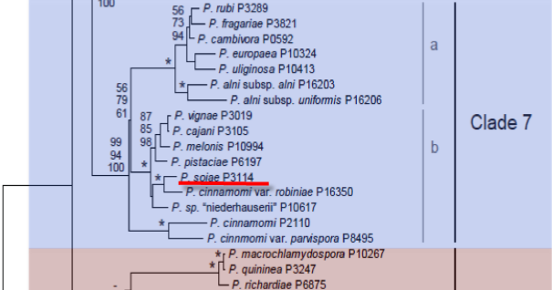
Genus wide phylogeny for Phytophthora using four mitochondrial loci (cox2, nad9, rps10 and secY; 2,373 nucleotides). Maximum likelihood branch lengths shown. Numbers on nodes represent bootstrap support values for maximum likelihood (top), maximum parsimony (middle) and Bayesian posterior probabilities as percentages (bottom). Nodes receiving significant support (>95%) in all analysis are marked with an asterisk (*). Scale bar indicates number of substitutions per site.(Martin, Blair and Coffey, unpublished).

[ Click the tree to enlarge it. ]
Phytophthora sojae Kaufm. & Gerd. 1958 (Oomycetes, Pythiales) Phytophthora megasperma var. sojae (Kauffm. & Gerd.) A.A. Hildebrand 1959
= Phytophthora megasperma f. sp. glycines T.L. Kuan & Erwin 1980 Note: Corrected from glycinea.
Variant spelling Phytophthora megasperma f. sp. glycinea T.L. Kuan & Erwin 1980 Note: Original spelling.
= Phytophthora sojae f. sp. glycines Faris et al. 1989 Note: Corrected from glycinea.
Notes: Previous to 1991, this species was commonly reported under the name Phytophthora megasperma.
Distribution: Australia, North America (Canada, USA). Also reported from South America (Chile), Asia (Korea, China) and New Zealand.
Substrate: Roots, stems.
Disease Note: Root and stem rot of soybeans; seedling wilt, seedling blight of lupines.
Host: Glycine max (soybeans), Lupinus spp. (lupine) (Fabaceae); also reported from six other genera in five families.
Supporting Literature:
Erwin, D.C., and Ribeiro, O.K. 1996. Phytophthora Diseases Worldwide. APS Press, St. Paul, Minnesota, 562 pages.
Faris, M.A., Sabo, F.E., Barr, D.J.S., and Lin, C.S. 1989. The systematics of Phytophthora sojae and P. megasperma. Canad. J. Bot. 67: 1442-1447
Hansen, E.M., and Maxwell, D.P. 1991. Species of the Phytophthora megasperma complex. Mycologia 83: 376-381
Kroon, L.P.N.M., Bakker, F.T., van den Bosch, G.B.M., Bonants, P.J.M., and Flier, W.G. 2004. Phylogenetic analysis of Phytophthora species based on mitochondrial and nuclear DNS sequences. Fungal Genet. Biol. 41: 766-782
Updated on Jun 12, 2006
P. sojae is classified in group V (Stamps et al. 1990). Morphology is shown in Figure 1 and Figure 2. See Tables 4.2 and 4.3 in Phytophthora Diseases Worldwide (Erwin and Ribeiro 1996) for tabular keys.
1. Sporangia
Sporangia are nonpapillate and ovoid, ellipsoid, and sometimes obpyriform. Sporangia range in size from 23.3 to 88.8 µm x 16.6 to 51.8 µm, average 58 x 38.3 (Kaufmann and Gerdemann 1958) (Figure 1 and Figure 2E-J). Sporangiophores are simple and undifferentiated. Sporangia are formed both sympodially and by internal and external proliferation (Hildebrand 1959).
2. Hyphal Swellings
Intercalary hyphal swellings, which are spherical to irregular in shape, occur especially in aqueous culture.
3. Chlamydospores
Chlamydospores are not mentioned in the original description by Kaufmann and Gerdemann (1958), but Hildebrand (1959) showed a photomicrograph of a chlamydospore (Figure 1) and stated that chlamydospores conformed to the definition of Blackwell (1949). They were similar in size to oogonia. Schmitthenner in Sinclair and Backman (1989) stated that chlamydospores occurred, but he did not give dimensions.
4. Sex Organs
P. sojae is homothallic. Oospores are formed in infected root tissue of susceptible and tolerant cultivars (Slusher and Sinclair 1973). Oogonia are spherical to subspherical and vary from 29.4 to 45.7, average 36.3 µm (Hildebrand 1959); 19.2 to 38.3, average 31 µm (Kaufmann and Gerdemann 1958); and 32 to 38 µm (Faris et al. 1989). Antheridia are mostly paragynous, but some are amphigynous (Hildebrand 1959; Faris et al. 1989). The diameters of oospores range from 19.2 to 38.3 µm (average 31.4 µm) (Kaufmann and Gerdemann 1958). Oospores nearly fill the oogonium (plerotic). Oospores germinate at relatively high percentages (Erwin and McCormick 1971; Schechter and Gray 1987; Bhat et al. 1990, 1992, 1993; Bhat 1991; Bhat and Schmitthenner 1992, 1993a, b). Figure 3.6 in Phytophthora Diseases Worldwide (Erwin and Ribeiro 1996) shows the progress of germinating oospores at different stages (Erwin and McCormick 1971). Figure 3.8 in Phytophthora Diseases Worldwide (Erwin and Ribeiro 1996) shows that after two nuclei fuse and the inner wall of the oospore erodes, the oospore swells and germinates (Jiang et al. 1989).
5. Growth Temperatures
Reports of optimum temperatures for growth include 25oC (Hildebrand 1959; Faris et al. 1989) and 20oC (Kaufmann and Gerdemann 1958; Hansen and Maxwell 1991). No growth occurs at 5 and 35oC (Kaufmann and Gerdemann 1958), but slight growth was reported by Hansen and Maxwell (1991). According to Schmitthenner in Sinclair and Backman (1989), 25-28oC is optimum and 32-35oC is maximum for growth.
6. Growth Characteristics
P. sojae does not grow on potato-dextrose agar (Figure 2L) (Kaufmann and Gerdemann 1958; Hildebrand 1959; Kuan and Erwin 1980a); however, addition of sitosterol (30 µg/ml) or dilution of potato-dextrose agar to 1:10 increases growth to normal (Erwin et al. 1968).
7. Distinguishing Characteristics
Although P. sojae, P. medicaginis, and P. trifolii have been classified within the species P. megasperma as formae speciales and have been considered to be relatively similar, several features readily distinguish P. sojae from P. medicaginis. Because P. trifolii has been noted only in one area of Mississippi, its characteristics are less known (see Chapter 62 in Phytophthora Diseases Worldwide [Erwin and Ribeiro 1996]). Table 1 is a compilation of physiological and morphological features that distinguish P. sojae from P. medicaginis (see also Nygaard et al. [1989] and Chapter 44 on P. medicaginis).
[[PAPER:2148|1]]
Nomenclature information was provided by the the Systematic Botany and Mycology Laboratory in USDA-ARS.
Isolate list