
Phytophthora has been rebuilt to fix security-related problems and to restore GIS tools. These tools allow users to visualize the geospatial, temporal, and environmental contexts of Phytophthora discoveries. The next phase is to update species information and add data derived from large-scale surveys. If you have suggestions and requests to make the database better, please contact Seogchan Kang (sxk55@psu.edu).
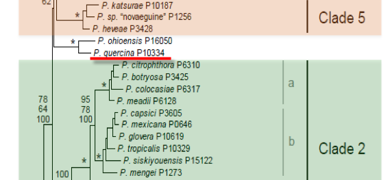
Genus wide phylogeny for Phytophthora using four mitochondrial loci (cox2, nad9, rps10 and secY; 2,373 nucleotides). Maximum likelihood branch lengths shown. Numbers on nodes represent bootstrap support values for maximum likelihood (top), maximum parsimony (middle) and Bayesian posterior probabilities as percentages (bottom). Nodes receiving significant support (>95%) in all analysis are marked with an asterisk (*). Scale bar indicates number of substitutions per site.(Martin, Blair and Coffey, unpublished).

[ Click the tree to enlarge it. ]
Phytophthora quercina T. Jung 1999 (Oomycetes, Pythiales)
Distribution: Asia (Turkey), Europe (Germany, Hungary, Italy).
Substrate: Roots, rhizosphere soil.
Disease Note: Implicated in oak decline, associated with various other Phytophthora spp.
Host: Quercus spp. (Fagaceae).
Supporting Literature:
Cooke, D.E.L., Jung, T., Williams, N.A., Schubert, R., Bahnweg, G., Oswald, W., and Duncan, J.M. 1999. Molecular evidence supports Phytophthora quercina as a distinct species. Mycol. Res. 103: 799-804
Jung, T., Cooke, D.E.L., Blaschke, H., Duncan, J.M., and Oswald, W. 1999. Phytophthora quercina sp. nov., causing root rot of European oaks. Mycol. Res. 103: 785-798
Updated on Jun 16, 2006
Morphology of spore structures is shown in Figure 1.
1. Sporangia
Produced in small numbers on solid agar and abundantly in liquid culture. Sporangia are noncaducous, papillate or occasionally bipapillate. Generally, terminal but occasionally intercalary. Sporangial shapes ranged from sub-globose, ovoid and obpyriform to ampuliform, banana or peanut-like distorted shapes. Sporangia with markedly curved apices are common. Lateral displacement of the papilla and short hyphal projections on sponrangia is present occasionally. Sporangiophores are simple or forming irregular lax sympodia, 1.5-5.8 µm (av.3.2 µm). Average dimensions of sporangia are 42.4 x 29.3 µm with a length-breadth ratio of 1.45.
2. Chlamydospores
Chlamydospores are occasionally produced, spherical, terminal or intercalary 17-35 µm
3. Sex Organs
P. quercina is homothallic. Oogonia are formed readily in single culture. Oogonial shapes ranged from spherical to ovoid and ellipsoid, 45% being markedly elongated as though assuming the shape of a host cell. On malt extract agar (MEA) they were on average 29.4 µm. Oospores shapes are spherical to ovoid (on average 26.6 µm), markedly aplerotic and thick-walled (average 2.5 µm). Older oospore walls often turned golden-yellow. Antheridia are hyaline, single, terminal, and spherical or club-shaped to irregular (average 14.8 x 9.5 µm). They are always paragynous, usually inserted near the oogonial stalk.
4. Growth Temperatures
It grows on V8 agar (V8A) at 5-27.5oC with an optimum near 25oC (radial growth rate 3.3 mm d-1). No growth occurred at 30oC.
5. Growth Characteristics in Culture Media
Colonies were uniform without distinct growth pattern on V8A and MEA (dome-shaped and fluffy), corn meal agar (CMA; sparse aerial mycelium), and potato dextrose agar (PDA; appressed densely-felty and dome-shaped).
6. Distinguishing Characteristics
P. quercina differed from Waterhouse\'s Group I species by its uniform, dome-shaped and cottonwool-like colony growth pattern on V8A and MEA, the frequent occurrence of sympodially branched primary hyphae, a high proportion of elongated, ellipsoid or ovoid oogonia, the absence of amphigynous antheridia and RAPD banding pattern (Cooke et al. 1999). P. quercina also is distinguished from the Group I species by its high variation in size and shape of the sporangia or large proportion of sporangia with cuverd apex, hyphal projections, lateral displacement of the papilla and lateral attachment to the sporangiophore.
Suggested to be involved in fine root mortality of oak trees and thus associated with oak decline.
Only detected from fine roots of oak species by direct plating on PARPNH selective medium and more easily via baiting of soil samples collected around the stem of an oak (Jung et al. 1996; Nechwatal et al. 2001). Never found be associated with bleeding cankers.
Management of infested material in nurseries. No management practices are available under forest settings.
[[PAPER:2386|1]]
[[PAPER:2340|1]]
[[PAPER:2345|1]]
[[PAPER:2387|1]]
[[PAPER:2388|1]]
[[PAPER:2389|1]]
[[PAPER:2346|1]]
[[PAPER:2390|1]]
[[PAPER:2383|1]]
[[PAPER:2391|1]]
[[PAPER:2392|1]]
Nomenclature information was provided by the the Systematic Botany and Mycology Laboratory in USDA-ARS.
This page was written by Yilmaz Balci at West Virginia University.