
Phytophthora has been rebuilt to fix security-related problems and to restore GIS tools. These tools allow users to visualize the geospatial, temporal, and environmental contexts of Phytophthora discoveries. The next phase is to update species information and add data derived from large-scale surveys. If you have suggestions and requests to make the database better, please contact Seogchan Kang (sxk55@psu.edu).
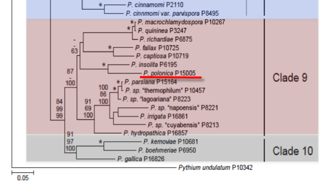
Genus wide phylogeny for Phytophthora using four mitochondrial loci (cox2, nad9, rps10 and secY; 2,373 nucleotides). Maximum likelihood branch lengths shown. Numbers on nodes represent bootstrap support values for maximum likelihood (top), maximum parsimony (middle) and Bayesian posterior probabilities as percentages (bottom). Nodes receiving significant support (>95%) in all analysis are marked with an asterisk (*). Scale bar indicates number of substitutions per site.(Martin, Blair and Coffey, unpublished).

[ Click the tree to enlarge it. ]
Phytophthora polonica Belbahri, Moralejo & Lefort 2006 (Oomycetes, Pythiales)
Distribution: Europe (Poland).
Substrate: Soil.
Disease Note: Slight disease on Alnus twigs in experiments.
Supporting Literature:
Maseko, B., Burgess, T. I. , Coutinho, T.A., and Wingfeld, M.J. 2007. Two new Phytophthora species from South African Eucalyptus plantations. Mycol. Res. 111: 1321-1338.
Updated on Mar 10, 2008
Phytophthora polonica Belbahri L, Moralejo E & Lefort F. was recovered from underneath declining alder trees in Poland. Phylogenetically it is a clade 9 species closely related to P. insolata.
1. Sporangia
Not observed on any culture media. None formed in water soil extract and only a few in gelatine solution. Borne on long nonbranching sporangiophores, mostly ovoid to ellipsoid, c. 52–67 X 32–44 µm, noncaducous, nonpapillate, proliferating internally, often nested or catenulate. Zoospores discharged through an exit pore 10–18 µm wide.
2. Chlamydospores
Abundant within 10 days on CA, CMA and in soil extract; spherical to subglobose or pyriform, average diameter 48.4 µm (ranging from 16 to 69 µm), moderately thin walled (1–2 µm), intercalary, lateral or terminal on short branches. In water, forming extensive networks of geniculate hyphae with lateral chlamydospores at the joints
3. Sex Organs
Abundant in single-zoospore isolates on CA after 1 week. Mostly borne on stalked branches, spherical to subglobose, smooth and thin-walled, 41.8 ±2.8 SD µm diameter. Oospores From aplerotic to nearly filling the oogonia, 38.1 ± 2.5 µm diameter, moderately thick walled (average 2.9 ± 0.8 µm) . A high proportion of aborted oogonia were seen. Antheridia Mostly clavate to irregularly shaped, less frequently spherical to barrel shaped, 16.2 ± 2.8 µm long X 13 ± 2.1 µm wide; borne on long stalks, mostly attached near the oogonial base, occasionally with hyphal extensions. Predominantly paragynous but sometimes amphigynous.
4. Growth Temperatures
Moderately slow on CA and CMA at 20° C and slow on PDA and MEA. Optimum temperature c. 30° C, minimum c. 5° C and maximum c. 38° C. In cultures on CA at 20° C contaminated by bacteria, growth was stimulated on the side facing the bacterial colonies.
5. Growth Characteristics in Culture
Main hyphae up to 8 µm wide. Colony pattern: aerial mycelium on CA appressed to limited, slightly stellate to rosaceous; concentric growth rings somewhat noticeable on the underside of the Petri dish, colony on CMA submerged and somewhat radiate, on MEA aerial mycelium appressed, fairly felty, markedly rosaceous, on PDA felty and broadly lobed, rosaceous. Hyphal swellings readily and abundantly formed, usually large (up to c. 50 µm long), single or more frequently catenulate, intercalary or lateral, or aggregated, inflated, toruloid, irregularly shaped to globose. Found either in agar or in soil water extract.
6. Distinguishing Characteristics
Phytophthora polonica exhibited a combination of unique morphological characters and distinctive nuclear and mitochondrial DNA sequences that easily enables distinction from other Phytophthora species. It belongs to group V of Waterhouse morphological scheme of classification by being homothallic with paragynous antheridia, and bearing nonpapillate sporangia with internal proliferation. Other Phytophthora spp. within group V include P. fragariae, P. rubi, P. humicola, P. insolita, P. medicaginis, P. megasperma, P. quininea, P. sojae and P. trifolii. Unlike P. polonica, both P. fragariae and P. rubi, as well as species within the ‘megasperma complex’ sensu Hansen et al. (1986) can be readily distinguished by having lower cardinal growth temperatures; P. humicola has unusually high optimal temperatures like P. polonica but does not form chlamydospores. Of those species included in clade 8 P. insolita superficially resembles P. polonica in its cardinal temperatures and colony pattern and in the formation of hyphal swellings and small chlamydospores, but it is easily distinguished by its parthenogenetic oospores, i.e. without attached antheridia; P. quininea differs by producing larger oogonia; P. richardiae is self-fertile and has a lower maximum growth temperature; and P. macrochlamydospora does not form sexual structures. It is noteworthy that all these species of clade 8 form survival structures such as hyphal swellings, chlamydospores or oospores in pure culture. By having hyphal swellings and chlamydospores, P. cinnamomi and P. lateralis could be confused with P. polonica; however, the first has higher colony growth rates and the second lower cardinal temperatures. Although occupying the same niche, P. alni and its subspecies are distinguished by their almost exclusive amphigynous antheridia and often nonsmooth oogonial walls.
Phytophthora polonica was recovered from soil around declining alder trees but in laboratory tests was slightly pathogenic to alder twigs and not pathogenic to trunks of several tree species.
Belbahri, L., Moralejo, E., Calmin, G., Oszako, T., Garcıa, J. A., Descals, E. and Lefort, F. 2006. Phytophthora polonica, a new species isolated from declining Alnus glutinosa stands in Poland. FEMS Microbiol. Lett. 261:165–174
This species page has been adapted from Belbahri et al. (2006)
Isolate list