
Phytophthora has been rebuilt to fix security-related problems and to restore GIS tools. These tools allow users to visualize the geospatial, temporal, and environmental contexts of Phytophthora discoveries. The next phase is to update species information and add data derived from large-scale surveys. If you have suggestions and requests to make the database better, please contact Seogchan Kang (sxk55@psu.edu).
Genus wide phylogeny for Phytophthora using four mitochondrial loci (cox2, nad9, rps10 and secY; 2,373 nucleotides). Maximum likelihood branch lengths shown. Numbers on nodes represent bootstrap support values for maximum likelihood (top), maximum parsimony (middle) and Bayesian posterior probabilities as percentages (bottom). Nodes receiving significant support (>95%) in all analysis are marked with an asterisk (*). Scale bar indicates number of substitutions per site.(Martin, Blair and Coffey, unpublished).

[ Click the tree to enlarge it. ]
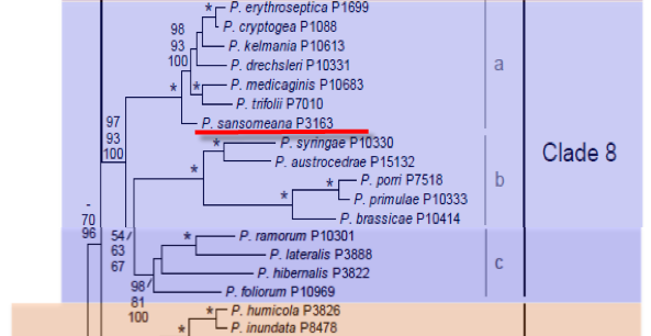
Phytophthora sansomeana E.M. Hansen & Reeser was first was characterized based on isolates from Douglas-fir seedlings in nurseries in Oregon and weeds (white clover, wild carrot, white cockle) in alfalfa fields in New York. They were labeled variously P. megasperma DF, DF1, D1, and Group 1 (see Hansen et al. 2009). More recently it was recognized that an unnamed Phytophthora species from soybeans in the Midwest was morphologically similar and shared growth and pathogenicity characteristics. Because P. sansomeana from soybean is better characterized ecologically, including pathogenicity, an Indiana soybean isolate was chosen as type. Phylogenetically it is in clade 8a basal to P. cryptogea and P. drechsleri. Isolates of P. sansomeana from Oregon, New York and the Midwestern United States carry ds RNA (see Hansen et al. 2009).
1. Sporangia
Sporangia are ovoid or obpyriform, nonpapillate, and average 56 µm long, (isolate means 54–59 µm) with a usual length to width ratio of 1.5–1.6 (Fig 1). Sporangia form in water, on long, unbranched or loosely sympodial sporangiophores. Proliferation is typically internal, occasionally lateral from beneath the sporangium.
2. Chlamydospores
Chlamydospores are not formed in agar.
3. Sex Organs
Phytophthora sansomeana is a homothallic species with predominately paragynous antheridia (Fig 1). Oogonia of soybean isolates average 39 µm diameter (isolate means 37–41 µm). Oogonia of isolates from other hosts are somewhat larger, averaging 40–45 µm diam.
4. Growth Temperatures
Growth of soybean isolates on CMA averages 7 mm/d at 25 C, and isolates from other hosts grow up to 10 mm/d, with optimum growth at 25–27 C and a maximum growth temperature above 35 C.
5. Growth Characteristics in Culture
Colonies exhibit a moderately aerial, broadly petaloid pattern on carrot agar (illustrated in Hansen et al 1986).
6. Distinguishing Characteristics
P. sansomeana is morphologically similar to the species in Waterhouse’s group 5, hence its early misidentification as P. megasperma. It is most readily distinguished from P. megasperma and similar species by the combined features of oogonium and sporangium size as well as its faster growth, pathogenic behavior and host preferences. It differs from other species in ITS clade 8 by its homothallism, distinguishing it from the heterothallic P. drechsleri and P. cryptogea, and by faster growth with different colony morphology than P. medicaginis and P. trifolii. It does not exhibit the host specific pathogenicity of the latter two species.
P. sansomeana has been reported to cause root diseases on soybean, Douglas fir and several weed species.
Hansen, E. M., Wilcox, W. F., Reeser, P. W., Sutton, W. 2009. Phytophthora rosacearum and P. sansomeana, new species segregated from the Phytophthora megasperma ‘‘complex’’. Mycologia 101:129-135.
Hansen, E. M., Brasier, C. M., Shaw, D. S., Hamm, P. B. 1986. The taxonomic structure of Phytophthora megasperma: evidence for emerging biological species groups. Trans. Br. Mycol. Soc. 87:557–573.
This species page was adapted from Hansen et al. (2009)
Isolate list