
Phytophthora has been rebuilt to fix security-related problems and to restore GIS tools. These tools allow users to visualize the geospatial, temporal, and environmental contexts of Phytophthora discoveries. The next phase is to update species information and add data derived from large-scale surveys. If you have suggestions and requests to make the database better, please contact Seogchan Kang (sxk55@psu.edu).
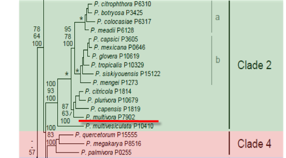
Genus wide phylogeny for Phytophthora using four mitochondrial loci (cox2, nad9, rps10 and secY; 2,373 nucleotides). Maximum likelihood branch lengths shown. Numbers on nodes represent bootstrap support values for maximum likelihood (top), maximum parsimony (middle) and Bayesian posterior probabilities as percentages (bottom). Nodes receiving significant support (>95%) in all analysis are marked with an asterisk (*). Scale bar indicates number of substitutions per site.(Martin, Blair and Coffey, unpublished).

[ Click the tree to enlarge it. ]
Phytophthora multivora P.M. Scott & T. Jung 2009 (Oomycetes, Pythiales)
Distribution: Widespread.
Host: Associated with several taxa.
Supporting Literature:
Scott, P.M., Burgess, T.I., Barber, P.A., Shearer, B.L., Stukely, M.J.C., Hardy, G.E.St.J., and Jung, T. 2009. Phytophthora multivora sp. nov., a new species recovered from declining Eucalyptus, Banksia, Agonis and other plant species in Western Australia. Persoonia 22: 1-13.
Updated on Nov 17, 2009
Phytophthora multivora P.M. Scott & T. Jung has been isolated in WA from natural forest and heath-land stands since the early 1980s from beneath dead and dying plants of 16 species from seven families (Burgess et al. 2009, Scott et al. 2009) but was misidentified as P. citricola. P. multivora is very widespread in south-west WA with a distribution similar to that known for P. cinnamomi. There is now evidence that in some sites it may be P. multivora and not P. cinnamomi that is responsible for tree mortality, while the latter is driving the collapse of whole ecosystems known as Phytophthora dieback. These findings may have direct implications for forest management and biosecurity. P. multivora has now been identified from other regions in Australia and, based on BLAST searches in GenBank, has been isolated in several additional countries (South Africa, Canada, Korea, Japan, Spain, Switzerland, Czech Republic, New Zealand, Hungary, USA). Several papers recent papers have compared species within the P. citricola complex. Sequence data for additional gene regions (EF1-α and NADH) were provided by Bezuidenhout et al. (2010).
1. Sporangia
Sporangia are rarely observed on solid agar but are produced abundantly in non-sterile soil extract. The majority of sporangia for all P. multivora isolates are formed between 7–12 h after flooding with soil extract. Little variation in sporangial shapes is observed between isolates. Sporangia are semipapillate and non-caducous and either ovoid (Fig. 1a), limoniform (Fig. 1b), ellipsoid or obpyriform sometimes with just a very shallow apical thickening. Sporangia with two or three papillae or distorted shapes are occasionally formed by all isolates (Fig. 1c,e). Sporangia are typically borne terminally but some are laterally attached or intercalary (Fig. 1d). External proliferation is regularly observed, either irregular or in lax or dense sympodia. The majority of sporangia of each isolate release zoospores between 15–20 h after flooding. The mean sporangial dimensions are 51.0 ± 10.4 × 30.0 ± 5.1 µm (overall range of 25–97 × 13–63 µm) with a length/breadth ratio of 1.7 ± 0.22 (overall range 1.3 – 3.3).
2. Chlamydospores
Chlamydospores not observed.
3. Sex Organs
P. multivora is homothallic. Gametangia are readily produced in single culture by all P. multivora isolates on V8A within 4 d. Oogonia of P. multivora are borne terminally, have smooth walls and are globose to slightly subglobose with a mean diameter of 26.5 ± 1.9 µm (overall range 19–37 µm and range of isolate means 25.5–27.8 µm). Oospores are globose (Fig. 2a-b) and nearly plerotic, oospore wall thickness 2.6 ± 0.5 µm (overall range 1.4–4.6 µm), oospore wall index 0.52 ± 0.07. Antheridia are obovoid, club-shaped or irregular, almost exclusively paragynous, diclinous and typically attached close to the oogonial stalk. Intercalary and amphigynous antheridia are only rarely observed. After 4 wk in V8A at 20° C, more than 90 % of oospores germinate directly.
4. Growth Temperatures
P. multivora grows on V8 agar (V8A) at 5-32.5° C with an optimum near 25° C (radial growth rate 4.7-6.1 mm/d). Generally no growth occurred at 32.5° C.
5. Growth Characteristics in Culture
On V8A it produces stellate growth patterns with a clearly delimited, submerged margin (Fig. 3a), on MEA it produces faintly stellate to dendroid patterns (Fig. 3b) and on PDA petaloid felty to fluffy colonies (Fig. 3c).
6. Distinguishing Characteristics
Compared with P. citricola, the sporangia are more uniform in shape and a higher proportion abort or germinate directly. P. multivora has significantly (P<0.05) thicker oospore walls (2.6 ± 0.5 µm, overall range 1.4–4.6 µm) than P. citricola (1.9 ± 0.3 µm, overall range 1.2–2.6). Due to the smaller oospore size of P. multivora the oospore wall index is significantly higher (P<0.0001) in P. multivora (0.52 ± 0.07) than in P. citricola (0.36 ± 0.05).
7. Type isolate
WESTERN AUSTRALIA, Yalgorup, from rhizosphere soil of declining Eucalyptus marginata, May 2007, P. Scott & T. Jung, holotype MURU 434 (dried culture on V8A, Herbarium of Murdoch University, Western Australia), culture ex-type WAC13201, CBS124094. rDNA ITS (FJ237521), coxI (FJ237508), β-tubulin (FJ665260).
Pathogenic toward Eucalyptus gomphocephala, E. marginata, Agonis flexuosa. Implicated in decline of Eucalyptus gomphocephala. Numerous other hosts including Banksia attenuata, B. grandis, B. littoralis, B. menziesii, B. prionotes, Bossiaea sp., Conospermum sp., Gastrolobium spinosum, Leucopogon verticillatus, Xanthorrhoea gracilis, Patersonia sp., Podocarpus drouyniana, Quercus petraea and Pinus radiata. Control attained by management of infested material in nurseries. No management practices are available under forest settings.
Commonly detected by baiting of rhizosphere soil, also isolated by direct plating of damaged roots or stem cankers on PARPNH selective medium.
Burgess, T. I., Webster, J. L., Ciampini, J. A., White, D., Hardy, G. E. S. J., Stukely, M. J. C.. 2009. Re-evaluation of Phytophthora species isolated during 30 years of vegetation health surveys in Western Australia using molecular techniques. Plant Disease 93: 215-223.
Bezuidenhout, C. M., Denman, S., Kirk, S. A., Botha, W. J., Mostert, L., McLeod, .A. 2010. Phytophthora taxa associated with cultivated Agathosma, with emphasis on the P. citricola complex and P. capensis sp. nov. Persoonia 25: 32-49.
Gallegly, M. E., Hong, C. 2008. Phytophthora: identifying species by morphology and DNA fingerprints. The American Phytopathological Society APS, St. Paul, Minnesota, USA.
Gallegly, M. E., Hong, C., Richardson, P., Kong, P. 2010. Phytophthora pini Leonian, a valid and distinct species. Phytopathology 100: S207.
Jung, T., Burgess, T. I.. 2009. Re-evaluation of Phytophthora citricola isolates from multiple woody hosts in Europe and North America reveals a new species, Phytophthora plurivora sp. nov. Persoonia 22: 95-110.
Moralejo, E., Pérez-Sierra, A. M., Álavez, L. A., Belbahri, L., Lefort, F., Descals, E. 2008. Multiple alien Phytophthora taxa discovered on diseased ornamental plants in Spain. Plant Pathology 57: 1–11.
Oudemans, P. V., Förster, H., Coffey, M. D.. 1994. Evidence for distinct isozyme subgroups within Phytophthora citricola and close relationships with P. capsici and P. citrophthora. Mycological Research 98: 189-199.
Scott, P. M., Burgess, T. I., Barber. P. A., Shearer, B. L., Stukely, M. J. C., Hardy, G. E. S. J., Jung, T. 2009. Phytophthora multivora sp. nov., a new species recovered from declining Eucalyptus, Banksia, Agonis and other plant species in Western Australia. Persoonia 22: 1-13.
Zea-Bonilla, .T, Martin-Sanchez, P. M., Hermoso, J. M., Carmona, M. P., Segundo, E., Perez-Jimenez, R. M. 2007. First report of Phytophthora citricola on Mangifera indica in Spain. Plant Pathology 556: 356–356.
This page was written by Treena Burgess and Thomas Jung, Centre for Phytophthora Science and Management, Murdoch University, Australia.
Isolate list