
Phytophthora has been rebuilt to fix security-related problems and to restore GIS tools. These tools allow users to visualize the geospatial, temporal, and environmental contexts of Phytophthora discoveries. The next phase is to update species information and add data derived from large-scale surveys. If you have suggestions and requests to make the database better, please contact Seogchan Kang (sxk55@psu.edu).
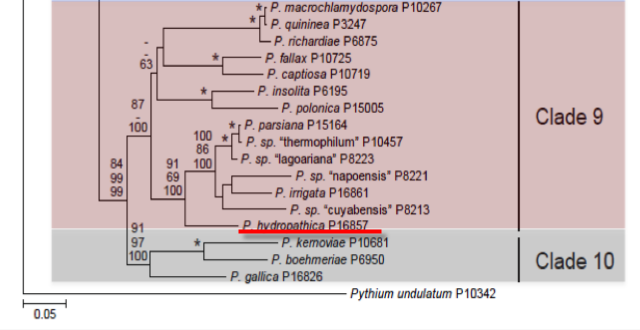
Genus wide phylogeny for Phytophthora using four mitochondrial loci (cox2, nad9, rps10 and secY; 2,373 nucleotides). Maximum likelihood branch lengths shown. Numbers on nodes represent bootstrap support values for maximum likelihood (top), maximum parsimony (middle) and Bayesian posterior probabilities as percentages (bottom). Nodes receiving significant support (>95%) in all analysis are marked with an asterisk (*). Scale bar indicates number of substitutions per site.(Martin, Blair and Coffey, unpublished).

[ Click the tree to enlarge it. ]
None
Phytophthora hydropathica Hong, C. and Gallegly, M. was initially recovered from irrigation reservoirs surveyed during the summer months. It is in clade 9 closely related to P. parsiana and P. irrigata.
1. Sporangia
Sporangia are produced on lima bean agar discs within 6 hr after they are placed in soil extract under lights, and the sporangia release zoospores within 2 hr after the sporangia are formed. Nesting and internal proliferation is very common. Sporangial shape varies from almost spherical, to ovoid and obpyriform. The sporangia are nonpapillate and noncaducous. The majority of sporangia tend toward being obpyriform. Their size is 43.3– 60.0 X 33.3–46.7 µm (average 55.3 X 38.9 µm). The sporangia form mostly on long sporangiophores. Hyphal swellings in hemp-seed agar are irregular in shape but often are obovate in clusters or at the end of hyphae. In water minute hyphal swellings are often catenulate.
2. Chlamydospores
Chlamydospores are terminal on short pedicels off the main hyphae and at the end of long hyphae. These thin-walled chlamydospores usually have dense protoplasm and are about 37 µmin diameter.
3. Sex Organs
Heterothallic. Small numbers of sexual bodies formed in P. hydropathica hemp-seed agar cultures beneath a polycarbonate membrane with a disc of A2 P. cinnamomi in hemp-seed agar on top; they were not formed with the A1 P. cinnamomi. The presence of sexual bodies beneath the A2, but not the A1 P. cinnamomi, indicates that all the P. hydropathica isolates tested were of the A1 mating type. Oogonia are about 43 µm in diameter, oospores 38 µm, and oospore walls about 2.2 µm thick. Antheridia are round and about 16 µm both tangential and perpendicular to the oogonia walls. Oospores are plerotic and the sexual bodies are golden in color in hemp-seed agar.
4. Growth Temperatures
The optimum temperature for colony growth was 30° C, whereas the maximum was 40° C, at which a trace of growth occurred. No growth occurred at 5° C during the first 2 days. Daily colony growth rates at 30° C ranged from 13.0 to 14.3 mm.
5. Growth Characteristics in Culture
The width of mycelium in some cultures seems relatively narrow, 3–5 µm. Colony pattern on PDA is petaloid.
6. Distinguishing Characteristics
Phytophthora hydropathica can be distinguished from the 39 other nonpapillate Phytophthora species by sexual pattern, morphology and growth temperature maximum. It is heterothallic and thus is easily separated from the 24 homothallic Phytophthora species that produce oospores in host tissue or agar media. There are three species (P. drechsleri, P. inundata and P. melonis) in the morphological key and two newly described species, P. irrigata and P. parsiana, that are heterothallic, nonpapillate and grow well at 35° C. Phytophthora hydropathica is a sixth species to be added to this group. It differs from P. drechsleri, P. inundata and P. melonis by having much larger oogonia and oospores which are golden in color. Also, sporangia differ by being mostly obpyriform and ovoid, whereas those of the other three species produce mainly ellipsoid sporangia, and P. hydropathica produces fully plerotic oospores, whereas the other three produce aplerotic to slightly aplerotic oospores. In contrast to P. irrigata, P. hydropathica produces chlamydospores and obovate hyphal swellings. Phytophthora parsiana and P. hydropathica are morphologically similar, but P. hydropathica has much larger oogonia and oospores, a higher temperature maximum, a faster growth rate, and different shapes of sporangia and hyphal swellings.
Phytophthora hydropathica causes leaf and stem necrosis on Rhododendron catawbiense and collar rot of Kalmia latifolia. It also causes seedling damping-off of cucumber (Cucumis sativus cv. Orient Express) and root rots of azaleas (Rhododendron Kurume hybrid cv. Hershey’s Red). It also infected the roots of dusty miller (Senecio bicolor subsp. cineraria cv. Silver Dust), tomato (Solanum lycopersicum cv. Homestead) and pepper (Capsicum annuum) plants. In addition, it can enter through wounds and cause fruit rot of peppers and tomatoes.
Hong, C. X., Gallegly, M. E., Richardson, P. A., Kong, P., Moorman, G. W., Lea-Cox, J. D. and Ross, D. S. (2010) Phytophthora hydropathica, a new pathogen identified from irrigation water, Rhododendron catawbiense and Kalmia latifolia. Plant Pathology 59:913-921.
This species page was adapted from Hong et al. (2010).
Isolate list