
Phytophthora has been rebuilt to fix security-related problems and to restore GIS tools. These tools allow users to visualize the geospatial, temporal, and environmental contexts of Phytophthora discoveries. The next phase is to update species information and add data derived from large-scale surveys. If you have suggestions and requests to make the database better, please contact Seogchan Kang (sxk55@psu.edu).
Genus wide phylogeny for Phytophthora using four mitochondrial loci (cox2, nad9, rps10 and secY; 2,373 nucleotides). Maximum likelihood branch lengths shown. Numbers on nodes represent bootstrap support values for maximum likelihood (top), maximum parsimony (middle) and Bayesian posterior probabilities as percentages (bottom). Nodes receiving significant support (>95%) in all analysis are marked with an asterisk (*). Scale bar indicates number of substitutions per site.(Martin, Blair and Coffey, unpublished).

[ Click the tree to enlarge it. ]
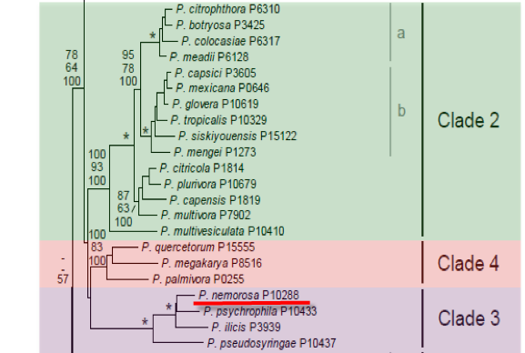
Phytophthora nemorosa E. M. Hansen and Reeser was recovered from bole cankers and leaf lesions of several tree species in California. Phylogenetic analysis places P. nemorosa in clade 3 closely related to P. psychrophila.
1. Sporangia
Sporangia formed in water are typically ovoid, scarcely semi-papillate, rarely bifurcate, appearing as terminal clusters on sympodially branched sporangiophores. Caducous sporangia averaged 51 µm in length (range 38-71 µm) and 37 µm in breadth (range 28 - 50 µm). Length:breadth ratios range from 1.3 to 1.5 (average 1.4). Pedicel lengths average 10 µm (range <5-20 µm), and apical plugs average 9 µm wide.
2. Chlamydospores
Only observed in older water cultures, mostly intercalary and occasionally terminal, 25.8 - 39.4 µm in diameter.
3. Sex Organs
Homothallic, producing terminal oogonia after 3 to 4 weeks in single culture on CMAβ. Oogonia average 33 µm (range 23 - 40 µm). Antheridia are amphigynous, approximately 13 x 13 µm (range 9-19 x 10-15 µm). Oospores are slightly aplerotic, averaging 29 µm (range form 19-35 µm).
4. Growth Temperatures
Optimum approximately 15° C with little or no growth above 20° C.
5. Growth Characteristics in Culture
Colony growth on CMAβ typically exhibit small blistered hyphal swellings.
6. Distinguishing Characteristics
Based on morphological features P. nemorosa is in Waterhouse Group IV. Three species in this group are homothallic with amphigynous antheridia and produce caducous semipapillate sporangia. P. hibernalis has longer pedicels and is primarily a pathogen of citrus while P. ilicis and P. phaseoli have smaller oogonia than P. nemorosa. P. nemorosa can be differentiated from the other clade 3 species based on morphology. It has smaller oogonia than P. psychrophila (33 vs 37 µm) as well as slightly rounder sporangia and a distinctly patterned growth on media. P. pseudosyringae and P. quercina can be differentiated based on their predominantly paragynous antheridia.
Phytophthora nemorosa and P. ramorum have a similar host range and symptomology, they can be differentiated by their growth in culture (P. nemorosa grows more slowly, has a lower temperature optimum; 15° C vs 20° C) as well as morphological features (P. nemorosa is homothallic).
Phytophthora nemorosa has been recovered from necrotic lesions of Umbellularia californica, Arctostaphylos spp., and Sequoia sempervirens. It has also been recovered from bark cankers of Lithocarpus densiflorus and Quercus agrifolia.
Hansen, E. M., Reeser, P., Davidson, J. M., Garbelotto, M., Ivors, K., Douhan, L., Rizzo, D. M. 2003. Phytophthora nemorosa, a new species causing cankers and leaf blight of forest trees in California and Oregon, USA. Mycotaxon 88: 129–138.
Nomenclature information was provided by the the Systematic Botany and Mycology Laboratory in USDA-ARS. This species page was adapted from Hansen et al.(2003)
Isolate list