
Phytophthora has been rebuilt to fix security-related problems and to restore GIS tools. These tools allow users to visualize the geospatial, temporal, and environmental contexts of Phytophthora discoveries. The next phase is to update species information and add data derived from large-scale surveys. If you have suggestions and requests to make the database better, please contact Seogchan Kang (sxk55@psu.edu).
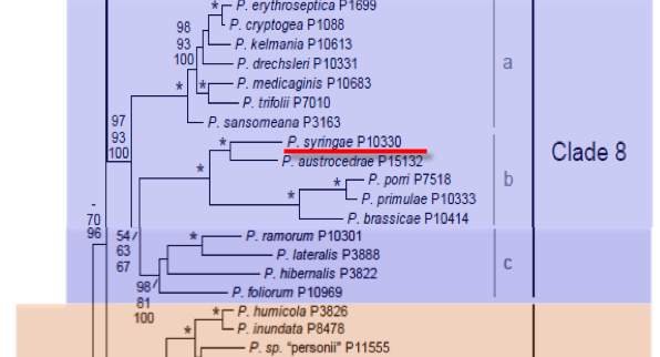
Genus wide phylogeny for Phytophthora using four mitochondrial loci (cox2, nad9, rps10 and secY; 2,373 nucleotides). Maximum likelihood branch lengths shown. Numbers on nodes represent bootstrap support values for maximum likelihood (top), maximum parsimony (middle) and Bayesian posterior probabilities as percentages (bottom). Nodes receiving significant support (>95%) in all analysis are marked with an asterisk (*). Scale bar indicates number of substitutions per site.(Martin, Blair and Coffey, unpublished).

[ Click the tree to enlarge it. ]
Phytophthora syringae (Berk.) Kleb. 1909 (Oomycetes, Pythiales) Ovularia syringae Berk. 1881Phytophthora cactorum subvar. syringae (Berk.) Sarej. 1936Nozemia syringae (Berk.) Pethybr. 1913Phloeophthora syringae (Berk.) Kleb. 1906 Note: Klebahn published Phloeophthora syringae as a new species, without reference to Ovularia syringae, but under Art. 33.2 it must instead be considered a new combination based on Ovularia syringae Berk..
= Pythiomorpha fischeriana Höhnk 1936 Note: Synonymy based on Erwin & Ribeiro (1996).
Phytophthora fischeriana (Höhnk) Sparrow 1960
Notes: Klebahn described this species in 1906 as a new species; according to Tucker (1931) it was previously described by Berkeley in 1881 as Ovularia syringae (misapplied to an anamorphic ascomycete genus). As a result the author citation should be (Berk.) Kleb. not (Kleb.) Kleb. as is frequently seen in the literature. Phytophthora hibernalis was considered by Tucker (1931) to be synonymous with Phytophthora syringae but they are now considered distinct species (Ho & Jong 1993). The name Phytophthora syringae has been applied to a root and collar rot of deciduous trees in Europe, which has now been described as a distinct species Phytophthora pseudosyringae (Jung et al. 2003).
Distribution: Africa (Morocco South Africa); Asia (Korea) Australasia; Europe; North America (Canada, USA); South America (Argentina, Brazil).
Substrate: Roots, shoots, twigs, trunks, leaves, fruits.
Disease Note: Twig blight, fruit rot, root rot, gummosis of citrus, stem canker, wilt, crown rot, collar rot; downy mildew, leaf spot, and shoot dieback of lilac.
Host: 29 genera in 14 families, including Syringa vulgaris (lilac, Oleaceae) and Rosaceae.
Supporting Literature:
Erwin, D.C., and Ribeiro, O.K. 1996. Phytophthora Diseases Worldwide. APS Press, St. Paul, Minnesota, 562 pages.
Ho, H.H., and Jong, S.C. 1993. Phytophthora hibernalis and P. syringae. Mycotaxon 47: 439-460
Jung, T., Nechwatal, J., Cooke, D.E.L., Hartmann, G.C., Blaschke, M., Oswald, W.F., Duncan, J.M., and Delatour, C. 2003. Phytophthora pseudosyringae sp. nov., a new species causing root and collar rot of deciduous tree species in Europe. Mycol. Res. 107: 772-789
Tucker, C.M. 1931. Taxonomy of the genus Phytophthora de Bary. Univ. Missouri Agric. Exp. Sta. Bull. 153: 1-208
Waterhouse, G.M., and Waterston, J.M. 1964. Phytophthora syringae. C.M.I. Descript. Pathog. Fungi Bact. 32: 1-2
Updated on Jun 07, 2006
P. syringae is classified in group III (Stamps et al. 1990). A summary is given by Waterhouse and Waterston (1964b). See Tables 4.2 and 4.3 in Phytophthora Diseases Worldwide (Erwin and Ribeiro 1996) for tabular keys. Morphology is shown in Figure 1.
1. Sporangia
Sporangia are broadly ovoid or obpyriform, semipapillate, and persistent and form in succession from a single sporangium. Dimensions are given in Table 1. The length-breadth ratio varies from 1.32:1 to 1.85:1. Sporangiophores form in a close monochasial sympodium with intercalary swellings.
2. Hyphal Swellings
Hyphal swellings are rounded or angular and often in chains (catenulate) and are occasionally delimited by septa. They often germinate by production of slender hyphae.
3. Chlamydospores
Chlamydospores are not produced according to Waterhouse and Waterston (1964b); however, Stamps et al. (1990) report chlamydospores 25 µm in diameter. Ho and Jong (1993) did not report chlamydospores.
4. Sex Organs
P. syringae is homothallic. Oogonia form abundantly in culture and host tissues; antheridia are predominately paragynous and occasionally amphigynous; and oospores fill the oogonium (plerotic). Dimensions of oogonia and oospores from different reports are given in Table 1. Oogonium diameter averages 33 µm, and oospore diameter averages about 30 µm. Germination of oospores in vitro was described by D. C. Harris and Cole (1982).
5. Growth Temperatures
The minimum temperature for growth is <5oC, the optimum is 15 to 20oC, and the maximum is 23 to 25oC.
6. Distinguishing Characteristics
Hyphae of old cultures are often knotty, vesicular, and coiled. The coiling of hyphae is similar to that observed in P. porri and P. primulae. P. syringae was erroneously considered by Tucker (1931) to be conspecific with P. hibernalis. These two species have often been confused. P. syringae produces noncaducous, ellipsoid sporangia and predominantly paragynous antheridia. P. hibernalis produces caducous, ovoid, obpyriform to elongated sporangia (larger than those of P. syringae) characterized by long pedicels (23–73 µm) and both paragynous and amphigynous antheridia in varying proportions (Ho and Jong 1993). Ho and Jong (1993) review the examples in the published literature where misidentification has led to confusion, e.g., Nadel-Schiffman (1947), who was corrected by Kouyeas and Chitzanidis (1968). Ho and Jong (1993) state that an N37 isolate from California labeled P. hibernalis actually produced amphigynous antheridia, indicating that it was probably P. syringae. Other citations in which P. hibernalis was identified as P. syringae include Wager (1942b) and Frezzi (1950). P. syringae differs from P. porri by its lower maximum temperature for growth and lack of chlamydospores. See Ho and Jong (1993) for a comparative study of P. syringae and P. hibernalis.
Nomenclature information was provided by the the Systematic Botany and Mycology Laboratory in USDA-ARS.
Isolate list