
Phytophthora has been rebuilt to fix security-related problems and to restore GIS tools. These tools allow users to visualize the geospatial, temporal, and environmental contexts of Phytophthora discoveries. The next phase is to update species information and add data derived from large-scale surveys. If you have suggestions and requests to make the database better, please contact Seogchan Kang (sxk55@psu.edu).
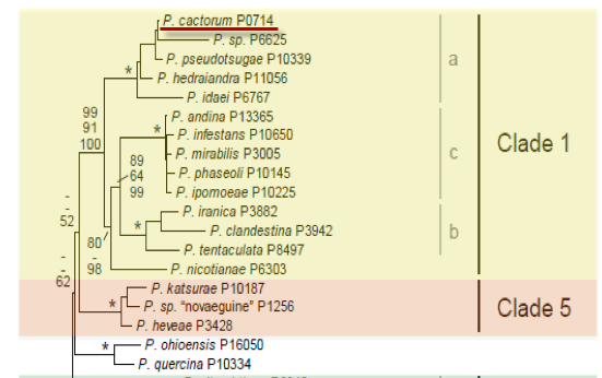
Genus wide phylogeny for Phytophthora using four mitochondrial loci (cox2, nad9, rps10 and secY; 2,373 nucleotides). Maximum likelihood branch lengths shown. Numbers on nodes represent bootstrap support values for maximum likelihood (top), maximum parsimony (middle) and Bayesian posterior probabilities as percentages (bottom). Nodes receiving significant support (>95%) in all analysis are marked with an asterisk (*). Scale bar indicates number of substitutions per site.(Martin, Blair and Coffey, unpublished).

[ Click the tree to enlarge it. ]
Phytophthora cactorum (Lebert & Cohn) J. Schr?. 1886 (Oomycetes, Pythiales) Peronospora cactorum Lebert & Cohn 1870Phytophthora cactorum var. cactorum (Lebert & Cohn) J. Schr?. 1886 Note: Other varieties are now considered synonyms of other species. Type variety not needed.Phytophthora omnivora de Bary 1881 Note: Illegitimate, superfluous name for P. cactorum (Waterhouse 1963).
= Peronospora fagi R. Hartig 1876
Phytophthora fagi (R. Hartig) R. Hartig 1879 Note: Sometimes erroneously cited as P. fagi Rosenbaum, but Rosenbaum listed P. fagi (R. Hartig) R. Hartig.
= Phytophthora paeoniae D.C. Cooper & Ch. Porter 1928
= Peronospora sempervivi Schenk 1875
Notes: Tucker (1931) listed P. citricola and P. pini as synonyms, but P. citricola (=P. pini) is now considered a distinct species (e.g., Waterhouse 1963, Martin & Tooley 2003, Erwin & Ribeiro 1996).
Distribution: Cosmopolitan.
Substrate: Leaves, stems, fruits, roots.
Disease Note: Damping off of seedlings, fruit rot, leaf and stem rot, collar and crown rot, stem canker, root rot (Waterhouse & Waterston 1966).
Host: At least 154 genera of vascular plants in 54 families (Waterhouse & Waterston 1966).
Supporting Literature:
Erwin, D.C., and Ribeiro, O.K. 1996. Phytophthora Diseases Worldwide. APS Press, St. Paul, Minnesota, 562 pages.
Martin, F.N., and Tooley, P.W. 2003. Phylogenetic relationships among Phytophthora species inferred from sequence analysis of mitochondrially encoded cytochrome oxidase I and II genes. Mycologia 95: 269-284
Tucker, C.M. 1931. Taxonomy of the genus Phytophthora de Bary. Univ. Missouri Agric. Exp. Sta. Bull. 153: 1-208
Waterhouse, G.M. 1963. Key to the species of Phytophthora de Bary. Mycol. Pap. 92: 1-22
Waterhouse, G.M., and Waterston, J.M. 1966. Phytophthora cactorum. C.M.I. Descript. Pathog. Fungi Bact. 111: 1-2
Updated on Jun 02, 2006
Morphology of spore structures is shown in Figures 1 and 2. See Waterhouse and Waterston (1966a) for a description of P. cactorum, Table 1 and 2 for tabular keys, and Appendix 4.9 in Phytophthora Diseases Worldwide (Erwin and Ribeiro 1996) for a dichotomous key (Ho 1992; Ho et al. 1995).
1. Sporangia
Sporangia are distinctively papillate, normally borne terminally, and occasionally intercalary. Shapes of sporangia range from broadly ellipsoidal, obpyriform, or ovoid to spherical. The sporangia are caducous with short pedicels (less than 4 μm in length). Sporangiophores are normally simple or in a close or lax sympodium with sporangia often clustered (Figure 2). Dimensions of sporangia are 31.4  4.8 x 26.4 4.0 with a length-breadth ratio of 1.2 0.1 (Oudemans and Coffey 1991a). Data reported in several references are given in Table 1. Media for sporangia production are evaluated by Fortes and Pecknold (1981). Several methods are given in the Appendix of Chapter 2 in Phytophthora Diseases Worldwide (Erwin and Ribeiro 1996).
2. Chlamydospores
Chlamydospores are produced by some but not all isolates. The somewhat erratic production of chlamydospores by certain isolates in different reports (Waterhouse and Waterston 1966) may have been caused by unfavorable environmental conditions. Several isolates from different hosts, including ginseng (Panax quinquefolium L.), formed chlamydospores abundantly in V8 juice-calcium carbonate broth when incubated for 20 days at 4oC, but none were produced at 8, 12, 16, 20, or 32oC. A few developed at 24 and 28oC. Chlamydospores did not form in potato-dextrose broth or pea seed broth. Chlamydospores in mycelial mats formed when buried in potting mix soil at 4oC. Chlamydospores germinated well (60 to 80%) even when subjected to freezing at 23oC for 24 h (Darmano and Parke 1990). A majority of the chlamydospores produced are terminal, although occasionally some are intercalary. The two-layered, single-wall thickness is usually 1 to 1.5 μm. Diameters (25 to 39.7 μm) of chlamydospores recorded in different reports are given in Table 2.
3. Sex Organs
P. cactorum is homothallic. Most isolates produce oospores in culture media and in diseased plant tissue. Antheridia are paragynous and unicellular and nearly spherical to club-shaped and often occur in a knot of hyphae. Antheridia measure 8.5 to 21 x 12 to 21 μm (Frezzi 1950); 15 x 13 μm (Waterhouse 1963); 9 to 20 x 7 to 13 μm, average 13.1 x 10.1 μm (Ershad 1971); and 9 to 12 x 9 to 20 μm (Gerrettson-Cornell 1989). Oogonia are smooth-walled and usually hyaline. Oospores have been reported to be plerotic with an average wall thickness of 2 μm; however, Oudemans and Coffey (1991a) reported oospores were aplerotic. Dimensions of oogonia and oospores in different reports are given in Table 3. Germination of oospores by production of sporangia or mycelium has been reported many times. See Table 3.3 in Chapter 3 of Phytophthora Diseases Worldwide (Erwin and Ribeiro 1996). Germination in the field begins after a rest period during the winter at soil temperatures of about 7.5oC and then increases rapidly within 22 days to 82% as the soil temperature increases to 13oC (Braun and Nienhaus 1959).
4. Growth Temperatures
The minimum temperature for growth is 2oC, the optimum 2oC, and the maximum 3oC.
5. Growth Characteristics in Culture Media
Colonies are slightly radiate, compact without a definite border, and fluffy but not dense and have short aerial hyphae. See Chapter 4 of Phytophthora Diseases Worldwide (Erwin and Ribeiro 1996) for more information.
6. Distinguishing Characteristics
P. cactorum is distinguished from P. iranica and P. pseudotsugae by its production of caducous sporangia with short pedicels. P. iranica and P. pseudotsugae produce noncaducous sporangia. Some of the antheridia of P. clandestina are amphigynous and some paragynous, whereas those of P. cactorum are mostly paragynous. Oospores of P. cactorum are plerotic, but those of P. clandestina are aplerotic. Isozyme analysis and molecular characterization (mtDNA RFLP) of P. cactorum (Förster and Coffey 1991; Oudemans and Coffey 1991a) showed it to be highly uniform and distinct from other species.
A summary of diseases caused by P. cactorum is given by Gjaerum et al. (1988). Host range and distribution is summarized in Tables 4.
Infected apple trees are commonly found in poorly drained areas of the orchard where heavy, wet soils remain saturated for extended periods of time. Above-ground symptoms generally include reduced tree vigor and growth, yellowing or chlorosis of leaves, and eventual collapse or death of the tree. Infected trees may decline slowly over one or more years, or they may collapse and die rapidly after resuming growth in the spring. Rapid death of trees usually occurs following excessively wet periods.

Crown rot of apple showing typical below ground symptoms. The boundary of the necrosis is the graft union between the susceptible rootstock (in this case MM 104), and the resistant scion (Red Delicious). This brick red discoloration is pretty typical of Phytophthora crown rot; the line of demarcation between diseased and healthy tissue distinguishes Phytophthora root and crown rot from other causes of tree decline and collapse such as drowning or winter injury. (Photo credit: Turner Sutton, NC State University).
[[PAPER:2311|1]]
[[PAPER:2312|1]]
[[PAPER:2314|1]]
[[PAPER:2315|1]]