
Phytophthora has been rebuilt to fix security-related problems and to restore GIS tools. These tools allow users to visualize the geospatial, temporal, and environmental contexts of Phytophthora discoveries. The next phase is to update species information and add data derived from large-scale surveys. If you have suggestions and requests to make the database better, please contact Seogchan Kang (sxk55@psu.edu).
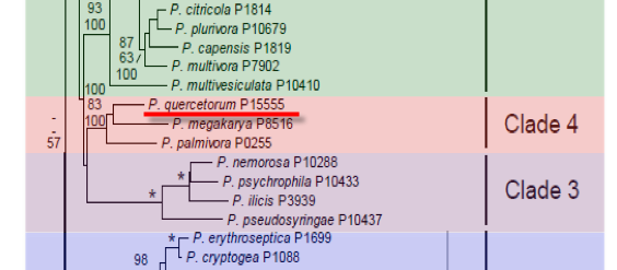
Genus wide phylogeny for Phytophthora using four mitochondrial loci (cox2, nad9, rps10 and secY; 2,373 nucleotides). Maximum likelihood branch lengths shown. Numbers on nodes represent bootstrap support values for maximum likelihood (top), maximum parsimony (middle) and Bayesian posterior probabilities as percentages (bottom). Nodes receiving significant support (>95%) in all analysis are marked with an asterisk (*). Scale bar indicates number of substitutions per site.(Martin, Blair and Coffey, unpublished).

[ Click the tree to enlarge it. ]
Phytophthora quercetorum Balci & Balci 2008 (Oomycetes, Pythiales)
P. quercetorum was first isolated during a soil survey in eastern and north-central US from oak forest rhizosphere soils (Balci et al. 2007).
Distribution: Throughout oak forests in Minnesota, Wisconsin, Ohio, Pennsylvania, Maryland and West Virginia in the United States (Figures 1 ).
Substrate: Roots, rhizosphere soil.
Disease Note: Implicated in oak decline, associated with Quercus macrocarpa, Q. rubra, Q. phellos. Pathogenic towards stem of seedlings and fine roots of various oak seedlings in artificial pathogenicity tests.
Host: Quercus spp. (Fagaceae).
Supporting Literature:
Balci Y, Balci S, Blair JE, Park SY, Kang S, MacDonald WL. 2008. Phytophthora quercetorum sp. nov., a novel species isolated from eastern and north-central U.S. oak forest soils. Mycological Research (in press)
Balci Y, Balci S, Eggers J, MacDonald WL, Juzwik J, Long RP, Gottschalk KW, 2007. Phytophthora species associated with forest soils in Eastern and North-central U.S. oak ecosystems. Plant Disease 91: 705-710.
Morphology of the species is shown in Figures 2 and Figures 3.
1. Sporangia
Sporangia were produced occasionally on solid agar substrate (V8A) and abundantly when agar plugs were immersed in soil extract water; with sympodial sporangiophores and no internal proliferation. Sporangia are non-caducous, papillate and occasionally bipapillate. Sporangia appear usually ovoid-elongated with common hyphal projections and frequently laterally attached. Displacement of papilla, globose and peanut-like distorted shapes were observed infrequently. In empty sporangia, a conspicuous basal plug was occasionally observed. Sporangial sizes (length x width) in eight isolates ranged from 17-40 x 14.5-32.5 µm (holotype 40.8 ± 5.8 x 30 ± 3.5 µm) with a length/width ratio of 1.4. Main hyphae was on average 5 µm thick (4-10 µm), with a coralloid branching pattern on V8A and markedly inflated in liquid culture.
2. Chlamydospores
Chlamydospores, which were rarely produced, were on average 30 ±2.9 µm.
3. Sex Organs
It is homothallic and produces gametangia abundantly on V8A within 5 days. Oogonia terminal at the main hyphae, globose, and predominantly turning yellow when mature. Mean oogonial diameter (eight isolates) was 31.4 ± 3 µm (holotype 31.8 ± 3 µm) ranging from 17 to 40 µm on V8A. The oogonial stalk predominantly bent or coiled, occasionally forming a curved tapering base. The tapering, elongated and coiled stalk as well as the markedly aplerotic oospores were more apparent in liquid culture. Oospores were always spherical, markedly aplerotic, and an average diameter (eight isolates) was 25.2 ± 2.3 µm (holotype 25.9 ± 2.2 µm), ranging from 14.5 to 32.5 µm. Average oospore wall thickness was 1.9 ± 0.7 µm. Average sizes of oogonia and oospores of the holotype strain on CMA were 31.1 µm and 24.4 µm, respectively. Antheridium was mostly lateral and sessile with short stalk, one per oogonium, attached near the stalk and rarely displaced, always paragynous, cylindrical or club-shaped, averaging 11.3 ± 2.2 x 9 ± 1.2 µm.
4. Growth Temperatures
Optimum temperature for growth on V8A (8 isolates) was ca. 22.5 °C, with upper temperature limit of ca. 32.5 °C (Figures 4). Radial growth rate at 22.5 °C in darkness ranged from 5.7-9.8 mm d-1 with an average of 7.5 mm d-1 (holotype 7.2 mm d-1).
5. Growth Characteristics in Culture Media
Colonies on CMA exhibit a striate pattern and are largely submerged, with no aerial mycelia; on V8A faintly stellate or radiate with restricted aerial mycelia, which disappear on older cultures; on MEA faintly petaloid; on PDA stoloniferous with a waxy appearance (Figures 5).
6. Distinguishing Characteristics
Being homothallic with paragynous antheridia and papillate sporangia, P. quercetorum falls in Group I of the Waterhouse classification based on morphological characteristics (Waterhouse 1963), which includes the following species: P. cactorum, P. clandestina, P. idaei, P. iranica, P. italica, P. pseudotsugae and P. tentaculata. Morphologically, P. quercetorum differs from P. cactorum by the absence of caducous and persistent ovoid or globose sporangia; from P. clandestina and P. iranica by absence of amphigynous antheridia and by sporangial features; from P. pseudotsugae and P. idaei by its markedly aplerotic, smaller oospores and production of distorted sporangia with hyphal projections; from P. tentaculata by the absence of arachnoid antheridia and abundant chlamydospores. It differs from P. italica by its average larger oogonia, oospores, antheridial sizes and its different sporangial features (Erwin & Ribeiro 1996). P. quercetorum can also be distinguished from P. hedraiandra (de Cock & Lèvesque 2004) by the absence of predominantly sessile antheridia and caducous sporangia and by different colony patterns on CMA and PDA.
Molecular analyses, however, suggest that P. quercetorum is more closely related to P. arecae, P. palmivora, P. megakarya, and P. quercina in Clade 4. P. quercetorum can be distinguished from P. aracae, P. palmivora, P. megakarya (Erwin & Ribeiro 1996) by being homothallic in culture and forming paragynous antheridia. It can be differentiated morphologically from P. quercina (Jung et al. 1999) by its spherical, non-elongated oogonial shapes and common coiled oogonial stalk, twice faster growth rate at the optimum temperature, and different colony types on V8A, MEA, PDA and CMA. The sporangia of P. quercetorum are similar to those of P. quercina, however, they are slightly shorter. P. quercetorum can be also separated from P. quercina by its coralloid branched hyphae and absence of protuberances.
Suspected to be involved in fine root mortality of oak trees and thus associated with oak decline.
Only isolated from soil via baiting of soil samples collected around the stem of oak (Balci et al. 2007, 2008). Never found associated with bleeding cankers of foliar necrosis.
Management of infested material in nurseries. No applied management practices are available under forest settings.
[[PAPER:2399|1]]
[[PAPER:2343|1]]
[[PAPER:2382|1]]
[[PAPER:2385|1]]
[[PAPER:2383|1]]
[[PAPER:2384|1]]
This page was prepared by Yilmaz Balci at West Virginia University.
Isolate list