
Phytophthora has been rebuilt to fix security-related problems and to restore GIS tools. These tools allow users to visualize the geospatial, temporal, and environmental contexts of Phytophthora discoveries. The next phase is to update species information and add data derived from large-scale surveys. If you have suggestions and requests to make the database better, please contact Seogchan Kang (sxk55@psu.edu).
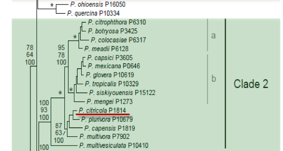
Genus wide phylogeny for Phytophthora using four mitochondrial loci (cox2, nad9, rps10 and secY; 2,373 nucleotides). Maximum likelihood branch lengths shown. Numbers on nodes represent bootstrap support values for maximum likelihood (top), maximum parsimony (middle) and Bayesian posterior probabilities as percentages (bottom). Nodes receiving significant support (>95%) in all analysis are marked with an asterisk (*). Scale bar indicates number of substitutions per site.(Martin, Blair and Coffey, unpublished).

[ Click the tree to enlarge it. ]
Phytophthora citricola Sawada 1927 (Oomycetes, Pythiales)
= Phytophthora cactorum var. applanata Chester 1932
= Phytophthora pini var. antirrhini Sundar. & T.S. Ramakr. 1928 Note: Synonymy based on Erwin & Ribeiro 1996.
Notes: Tucker (1931) treated P. citricola as a synonym of Phytophthora cactorum, but Waterhouse (1963) and Ho & Chang (1992) recognized it as a distinct species. Cooke et al. (2000) suggested that Phytophthora inflata is conspecific with Phytophthora citricola, but Kroon et al. (2004) showed that the two species differ in their mitochondrial DNA. Hong (2011) recently showed that P. pini, which had been considered to be a synonym of this name, was a distinct species.
Distribution: Cosmopolitan.
Substrate: Roots, fruits, stems, bark of trunks.
Disease Note: Brown rot of oranges, black root of hops, root rot and cane die-back of raspberry, die-back of rhododendron and basal rot of tomato seedlings (Waterhouse & Waterston 1966).
Host: 75 genera in 38 families.
Supporting Literature:
Cooke, D.E.L., Drenth, A., Duncan, J.M., Wagels, G., and Brasier, C.M. 2000. A molecular phylogeny of Phytophthora and related Oomycetes. Fungal Genet. Biol. 30: 17-32.
Erwin, D.C., and Ribeiro, O.K. 1996. Phytophthora Diseases Worldwide. APS Press, St. Paul, Minnesota, 562 pages.
Ho, H.H., and Chang, H.S. 1992. A re-evaluation of Phytophthora species described by K. Sawada in Taiwan. Mycotaxon 43: 297-316.
Hong, C., Gallegly, M.E., Richardson, P.A., and Kong, P. 2011. Phytophthora pini Leonian resurrected to distinct species status. Mycologia 103: 351-360.
Kroon, L.P.N.M., Bakker, F.T., van den Bosch, G.B.M., Bonants, P.J.M., and Flier, W.G. 2004. Phylogenetic analysis of Phytophthora species based on mitochondrial and nuclear DNS sequences. Fungal Genet. Biol. 41: 766-782.
Tucker, C.M. 1931. Taxonomy of the genus Phytophthora de Bary. Univ. Missouri Agric. Exp. Sta. Bull. 153: 1-208.
Waterhouse, G.M. 1963. Key to the species of Phytophthora de Bary. Mycol. Pap. 92: 1-22.
Waterhouse, G.M., and Waterston, J.M. 1966. Phytophthora citricola. C.M.I. Descr. Pathog. Fungi Bact. 114: 1-2.
Updated on Jul 20, 2011
P. citricola is classified in group III of Stamps et al. (1990). Morphology is shown in Figure 1 and Figure 2. See Tables 4.2 and 4.3 for tabular keys and Appendix 4.9 for a dichotomous key (Ho 1992) in Phytophthora Diseases Worldwide (Erwin and Ribeiro 1996). See Waterhouse (1957) and Zentmyer et al. (1974) for microphotographic exposition of sporangium and oospore morphology and Waterhouse and Waterston (1964d) for a general description.
1. Sporangia
Shapes of sporangia are highly variable (Figure 1 and Figure 2) and range from obovoid, obclavate, and obpyriform to slightly flattened on one side; occasionally deeply bifurcated with two apices; or irregularly shaped with three to four lobes. Zentmyer et al. (1974) show a wide range of variability in shapes. Sporangia are 30 to 75 μm long x 21 to 44 μm wide (average 47 x 34 μm), noncaducous, and persistent on the stalk. The septum is flush with the base of the sporangium. The papillae are wide and flat (semipapillate). Sporangiophores are simple or occasionally sympodial. A single terminal sporangium is sometimes borne on an unbranched sporangiophore.
2. Chlamydospores
Chlamydospores are rare (reported on oatmeal agar only).
3. Sex Organs
P. citricola is homothallic. Antheridia are paragynous (rarely amphigynous); oogonia are spherical and 18 to 35 μm in diameter, average 25.5 μm, sometimes tapering to a funnel-shaped base; oospores are spherical, almost plerotic, and 16 to 30 μm in diameter, average 22.0 μm.
4. Growth Temperatures
The minimum temperature for growth is 3oC; optimum is 25 to 28oC; and maximum is 31oC (Waterhouse 1963; Zentmyer et al. 1974).
5. Growth Characteristics in Culture Media
Avocado isolates produced radiate growth with pointed petallate zones that were more obvious on cornmeal agar than on V8 juice agar (Zentmyer et al. 1974).
6. Distinguishing Characteristics
Waterhouse\'s (1957) restudy of P. citricola in relation to reports by Sawada (1927), Chester (1932), and Ito and Tokunaga (1935) shows conclusively that P. citricola can be readily differentiated from P. cactorum var. applanata of Chester (1932). P. cactorum is conspicuously papillate [see Figure 11.1, Chapter 11 in Phytophthora Diseases Worldwide (Erwin and Ribeiro 1996)], whereas P. citricola has only a shallow but distinguishable thickening on the apex of sporangia (Figure 1 and Figure 2). Also, P. cactorum sporangia are shed in water (caducous with short pedicels; Figure 11.1 in Phytophthora Diseases Worldwide (Erwin and Ribeiro 1996), but those of P. citricola are not (Table 1). The oogonia of P. citricola and P. cactorum are very similar, but oogonia of P. cactorum are usually smaller and less pigmented than those of P. citricola. The radial growth of P. citricola is almost twice as rapid as that of P. cactorum. Other differences are given in Table 1.
6. Distinguishing Characteristics
On the basis of isozyme and mtDNA RFLP patterns, P. citricola isolates are divided into five subgroups (CIT1-IT5) (Oudemans and Coffey 1993). CIT1 is comprised of isolates from various Citrus species, including the Sawada (1927) type culture, an isolate previously known as P. pini (Leonian 1934), and an isolate identified as P. cactorum var. applanta (Chester 1932), now considered a synonym of P. citricola. Isolates in subgroups CIT1, CIT2, and CIT3 differ only at a few enzyme loci and are from diverse locations and hosts. Isolates in subgroup CIT4 are from South African plants, and CIT5 is composed of isolates pathogenic only to avocado (Zentmyer et al. 1974). The CIT5 isolates from avocado survive at lower temperatures (30oC) than isolates of the other groups (33C), and oogonia are consistently slightly smaller than those of the other groups. When cluster analyses were made, CIT5 clusered closer to P. capsici (from cocoa) and P. citrophthora than to the other CIT groups. The CIT5 group was considered to be a host-specific, genetically distinct group that could be regarded as a separate biological species (Forster and Coffey 1991; Oudemans and Coffey 1993). All of the CIT groups differ substantially from P. cactorum, thus further supporting the general conclusion that the P. cactorum var. applanta erected by Chester (1932) is not valid (Waterhouse 1957). Forster et al. (1990) also showed via mtDNA RFLP patterns that isolates of P. citricola are genetically diverse. Protein pattern analysis also showed diversity within P. citricola isolates (Erselius and de Vallavielle 1984). Isolates of CIT3 were shown to be more sensitive to metalaxyl than isolates of CIT1 (Ferrin and Kabashima 1991).
Host range and distribution is summarized in Table 2
Nechwatal, J., Schlenzig, A., Jung, T., Cooke, D.E.L., Duncan, J.M., Owald, W.F. 2001. A Combination of Baiting and PCR Techniques for the Detection of Phytophthora quercina and P. citricola in Soil Samples from Oak Stands. Forest Pathology 31: 85-97. Shew, H. D., and Benson, D. M. 1981. Fraser fir root rot induced by Phytophthora citricola. Plant Dis. 65:688-689.
Nomenclature information was provided by the the Systematic Botany and Mycology Laboratory in USDA-ARS.
Isolate list