
Phytophthora has been rebuilt to fix security-related problems and to restore GIS tools. These tools allow users to visualize the geospatial, temporal, and environmental contexts of Phytophthora discoveries. The next phase is to update species information and add data derived from large-scale surveys. If you have suggestions and requests to make the database better, please contact Seogchan Kang (sxk55@psu.edu).
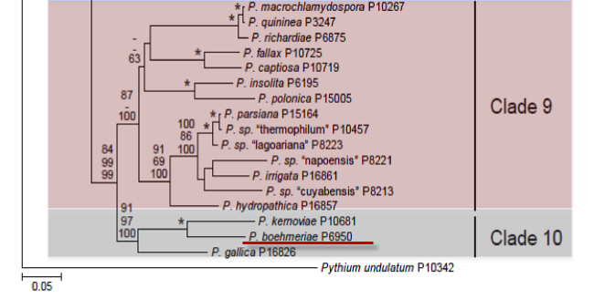
Genus wide phylogeny for Phytophthora using four mitochondrial loci (cox2, nad9, rps10 and secY; 2,373 nucleotides). Maximum likelihood branch lengths shown. Numbers on nodes represent bootstrap support values for maximum likelihood (top), maximum parsimony (middle) and Bayesian posterior probabilities as percentages (bottom). Nodes receiving significant support (>95%) in all analysis are marked with an asterisk (*). Scale bar indicates number of substitutions per site.(Martin, Blair and Coffey, unpublished).

[ Click the tree to enlarge it. ]
Phytophthora boehmeriae Sawada 1927 (Oomycetes, Pythiales)
Distribution: Africa (South Africa), Asia, Australia and Papua New Guinea, Europe (Greece), South America (Argentina).
Substrate: Fruits, leaves, roots, bolls.
Disease Note: Brown rot of citrus. Also leaf blight, root rot, cotton boll blight and rot (Erwin & Ribeiro 1996).
Host: Citrus spp. (Rutaceae) and various other plant families.
Supporting Literature:
Erwin, D.C., and Ribeiro, O.K. 1996. Phytophthora Diseases Worldwide. APS Press, St. Paul, Minnesota, 562 pages.
Ho, H.H., and Chang, H.S. 1992. A re-evaluation of Phytophthora species described by K. Sawada in Taiwan. Mycotaxon 43: 297-316.
Stamps, D.J. 1978. Phytophthora boehmeriae. C.M.I. Descr. Pathog. Fungi Bact. 591: 1-2.
Tucker, C.M. 1931. Taxonomy of the genus Phytophthora de Bary. Univ. Missouri Agric. Exp. Sta. Bull. 153: 1-208.
Waterhouse, G.M. 1963. Key to the species of Phytophthora de Bary. Mycol. Pap. 92: 1-22.
Updated on Jun 02, 2006
P. boehmeriae is classified in group II (Stamps et al. 1990). See Stamps (1978a) for a description, Tables 4.2 and 4.3 in Phytophthora Diseases Worldwide (Erwin and Ribeiro 1996) for tabular keys, and Ho (1992; Appendix 4.9) for a dichotomous key in Phytophthora Diseases Worldwide (Erwin and Ribeiro 1996). Morphology is shown in Figure 1.
1. Sporangia
Sporangia are papillate and spherical, ovoid, ellipsoidal, or obturbinate. The spores measure 28 to 69 x 20 to 51 μm, average 51.77 x 40.08 μm (Tucker 1931); 20 to 70 x 15 to 51 μm, average 44 x 32 μm (Frezzi 1950) (Table 2); 50 x 35 to 40 μm (Waterhouse 1963); 27 to 72 x 20 to 46 μm, average 49.9 x 34.5 μm (Katsura 1971); and 42 to 50 x 34 to 39 μm (Gerrettson-Cornell 1989). Length-breadth ratios reported are 1.25 to 1.4 (Waterhouse 1963) and 1.0 to 1.6 (Gerrettson-Cornell 1989). Sporangia are caducous with a pedicel length of 5.0 μm or less. Sporangiophores are sympodial.
2. Chlamydospores
Chlamydospores are produced infrequently. Diameters range from 26 to 51 μm, averaging 41.42 μm (Tucker 1931); 17 to 42 μm, averaging 29.5 μm (Frezzi 1950); 40.0 μm (Waterhouse 1963); and 30 to 41 μm (Gerrettson-Cornell 1989). Walls are 2 μm thick.
3. Sex Organs
P. boehmeriae is homothallic, and oospores form abundantly in host tissues. Antheridia are amphigynous and almost spherical in shape, often with a residual oil globule. Dimensions reported are 8 to 16 x 12.5 to 21 μm (Frezzi 1950); 14 to 21 x 13 μm (Waterhouse 1963); and 13 to 21 x 8 to 19 μm, average 14 to 17 x 14 to 16 μm (Gerrettson-Cornell 1989) (Table 4.3). Oogonia are smooth-walled and hyaline to yellow. Diameters are 21.7 to 31.7 μm, average 28 μm (Waterhouse 1963); and 19 to 41 μm, average 26 to 38 μm (Gerrettson-Cornell 1989). Oospores nearly fill the oogonium (plerotic). The wall is 2 μm or less in thickness. Oospore diameters are 20.9 to 27.6 μm, average 25.1 μm (Tucker 1931); 16 to 34 μm, average 27.5 μm (Frezzi 1950); average 24 μm (Waterhouse 1963); 9 to 12 x 9 to 14 μm (Katsura 1971); 16 to 35 μm, average 23 to 30 μm (Gerrettson-Cornell 1989) [Table 4.3, Phytophthora Diseases Worldwide (Erwin and Ribeiro 1996)].
4. Growth Temperatures
Minimum temperature for growth is 5 to 6oC, optimum is 25oC, and maximum is 32.5oC
5. Growth Characteristics in Culture Media
Colonies are uniform and compact with well-defined margins and dense aerial mycelia. Oospores, chlamydospores, and sporangia form abundantly in an agar medium in 7 to 10 days. P. boehmeriae is one of the most sensitive of all Phytophthora species to metalaxyl (linear growth on cornmeal agar was inhibited at 2 to 30 ng/ml). Because of its sensitivity, it has been used as a bioassay for metalaxyl in soil (A. M. Bailey and Coffey 1984–1986), plant tissue (Milgroom and Fry 1987), and in vitro (Chauhan and Singh 1987).
6. Distinguishing Characteristics
P. boehmeriae can be recognized by the production of abundant oogonia and oospores in single culture, amphigynous antheridia, and broadly ovoid to subspherical conspicuously papillate sporangia (Figure 1). P. boehmeriae differs from P. parasitica and P. citrophthora by production of oospores in single culture and from P. megasperma by production of papillate sporangia (Waterhouse 1963). Although 11 isolates of P. boehmeriae showed much genetic diversity by sozyme analysis, P. boehmeriae differed significantly from the morphologically similar species, P. katsurae and P. heveae Oudemans and Coffey (1991b).
Host range and distribution is summarized in Table 1.
Nomenclature information was provided by the the Systematic Botany and Mycology Laboratory in USDA-ARS.
Isolate list