
Phytophthora has been rebuilt to fix security-related problems and to restore GIS tools. These tools allow users to visualize the geospatial, temporal, and environmental contexts of Phytophthora discoveries. The next phase is to update species information and add data derived from large-scale surveys. If you have suggestions and requests to make the database better, please contact Seogchan Kang (sxk55@psu.edu).
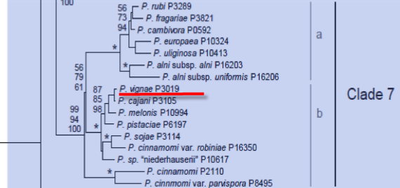
Genus wide phylogeny for Phytophthora using four mitochondrial loci (cox2, nad9, rps10 and secY; 2,373 nucleotides). Maximum likelihood branch lengths shown. Numbers on nodes represent bootstrap support values for maximum likelihood (top), maximum parsimony (middle) and Bayesian posterior probabilities as percentages (bottom). Nodes receiving significant support (>95%) in all analysis are marked with an asterisk (*). Scale bar indicates number of substitutions per site.(Martin, Blair and Coffey, unpublished).

[ Click the tree to enlarge it. ]
Phytophthora vignae Purss 1957 (Oomycetes, Pythiales)Phytophthora vignae f. sp. adzukicola S. Tsuchiya, Yanagawa & Ogoshi 1986 Note: Form species have no standing in the ICBN. This one applies to isolates that infect Vigna angularis. Phytophthora vignae f. sp. vignae S. Tsuchiya, Yanagawa & Ogoshi 1986 Note: Form species have no standing in the ICBN. This one applies to isolates that infect Vigna unguiculata. Notes: Tsuchiya et al. (1986) used the name Phaseolus radiatus var. aurea for adzuki bean. This bean is usually associated with the name Vigna angularis. Some consider Phaseolus radiatus var. aurea to be a synonym. Presumably, true adzuki bean was studied. Although the two form species of Phytophthora vignae may be distinct taxa, no one has formally recognized them as such. Distribution: Asia, Australia. Substrate: Stems, soil; also leaves, roots. Disease Note: Stem rot, leaf spot, may completely destroy root system. Host: Natural hosts: Vigna unguiculata (= Vigna sinensis, Fabaceae), Vigna angularis (=Phaseolus angularis, Fabaceae, sometimes referred to Phytophthora vignae f.sp. adzukicola). Malus domestica (apple, Rosaceae) fruits can be infected when inoculated. Reports from a few other taxa. Supporting Literature: Erwin, D.C., and Ribeiro, O.K. 1996. Phytophthora Diseases Worldwide. APS Press, St. Paul, Minnesota, 562 pages. Kroon, L.P.N.M., Bakker, F.T., van den Bosch, G.B.M., Bonants, P.J.M., and Flier, W.G. 2004. Phylogenetic analysis of Phytophthora species based on mitochondrial and nuclear DNS sequences. Fungal Genet. Biol. 41: 766-782. Tsuchiya, S., Yanagawa, M., and Ogoshi, A. 1986. Formae speciales differentiation of Phytophthora vignae isolates from cowpea and adzuki bean. Ann. Phytopathol. Soc. Japan 52: 577-584. Updated on Nov 21, 2008
P. vignae is classified in group VI (Stamps et al. 1990). See Tables 4.2 and 4.3 in Phytophthora Diseases Worldwide (Erwin and Ribeiro 1996) for tabular keys. The mycological description by Purss (1957) is summarized here. Morphology is shown in Figure 1.
1. Sporangia
Sporangia are variable in shape, ovoid to obpyriform, tapering toward the base; nonpapillate; persistent on the stalk; and proliferate both externally and internally. They are 24 to 72 μm long x 15 to 54 μm wide (average 48 x 27 μm) with a length-breadth ratio of 1.3:1 to 2.0:1 (average 1.8:1). Sporangia form in water or soil extract but do not form on solid culture media. They form on undifferentiated sporangiophores in aqueous culture, but on diseased tissues they form on monochasial or compound sympodia.
2. Hyphal Swellings
Hyphal swellings form abundantly and are large, spherical or angular, intercalary, and rarely terminal.
3. Chlamydospores
Chlamydospores are sparse in culture, terminal or intercalary, and measure 12 to 21 μm in diameter (average 17 μm).
4. Sex Organs
P. vignae is homothallic; oospores form abundantly in host tissues and in axenic culture; antheridia are variable in shape from spherical to ovate (average 16 x 15 μm) and amphigynous; oogonia are smooth walled and measure 24 to 46 μm in diameter (average 32 μm); oospores are markedly aplerotic (typically loose and eccentric within the oogonium) and measure 18 to 32 μm in diameter (average 26 μm); wall thickness is 3 μm.
Oospores of P. vignae were produced on V8 juice agar amended with sitosterol (30 mg/liter) at 20C and isolated free of mycelial debris by flotation and sedimentation. Oospores germinated readily on water agar to form nonpapillate sporangia. Germination was optimal at 25C. Oospore germination was as high as 70 to 90% at 20 to 30 days of age; but at 120 to 210 days of age, germination was reduced to less than 10% (Ishiguro and Ui 1981a). Oospore germination was reduced markedly by an increase in the osmotic potential of water agar with KCl or NaCl to -0.1 bar. An increase to -3.0 bars totally inhibited germination.
5. Growth Temperatures
The minimum temperature for growth is 10 to 12oC, optimum 28 to 30oC, and maximum 32oC, with no growth at 34.5oC.
Nomenclature information was provided by the the Systematic Botany and Mycology Laboratory in USDA-ARS.
Isolate list