
Phytophthora has been rebuilt to fix security-related problems and to restore GIS tools. These tools allow users to visualize the geospatial, temporal, and environmental contexts of Phytophthora discoveries. The next phase is to update species information and add data derived from large-scale surveys. If you have suggestions and requests to make the database better, please contact Seogchan Kang (sxk55@psu.edu).
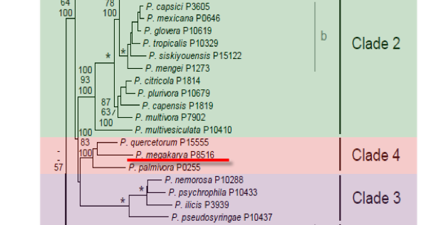
Genus wide phylogeny for Phytophthora using four mitochondrial loci (cox2, nad9, rps10 and secY; 2,373 nucleotides). Maximum likelihood branch lengths shown. Numbers on nodes represent bootstrap support values for maximum likelihood (top), maximum parsimony (middle) and Bayesian posterior probabilities as percentages (bottom). Nodes receiving significant support (>95%) in all analysis are marked with an asterisk (*). Scale bar indicates number of substitutions per site.(Martin, Blair and Coffey, unpublished).

[ Click the tree to enlarge it. ]
Phytophthora megakarya Brasier & M.J. Griffin 1979 (Oomycetes, Pythiales)
Notes: The species was previously reported under the name Phytophthora palmivora, S type. It closely resembles Phytophthora faberi (=Phytophthora palmivora var. palmivora); the name Phytophthora megakarya ex faberi has been suggested but is not currently in use (Erwin & Ribeiro 1996).
Distribution: Africa (Nigeria, Cameroon, Ghana, Fernando Po) (Erwin & Ribeiro 1996).
Substrate: Pods, seedlings, bark of trunks, possibly roots.
Disease Note: Pod rot, seedling blight, trunk canker. Inoculum does not survive in mummified pods, but survives in soil up to 18 months, possibly in infected roots (Erwin & Ribeiro 1996).
Host: Theobroma cacao (cacao, Sterculiaceae). Also reported from Irvingia sp. (Irvingiaceae).
Supporting Literature:
Erwin, D.C., and Ribeiro, O.K. 1996. Phytophthora Diseases Worldwide. APS Press, St. Paul, Minnesota, 562 pages.
Kroon, L.P.N.M., Bakker, F.T., van den Bosch, G.B.M., Bonants, P.J.M., and Flier, W.G. 2004. Phylogenetic analysis of Phytophthora species based on mitochondrial and nuclear DNS sequences. Fungal Genet. Biol. 41: 766-782
Stamps, D.J. 1985. Phytophthora megakarya. C.M.I. Descript. Pathog. Fungi Bact. 832: 1-2
Updated on Jun 12, 2006
P. megakarya is classified in group II (Stamps et al. 1990). A summary description of morphology and diseases caused is given by Stamps (1985b). Morphology is illustrated in Figure 1. See Tables 4.2 and 4.3 in Phytophthora Diseases Worldwide (Erwin and Ribeiro 1996) for tabular keys.
1. Sporangia
Sporangia are papillate and limoniform, obpyriform, or ellipsoid with rounded bases and are caducous with pedicels of intermediate length (10 to 30 µm). Size varies from 20 to 60 x 13 to 41 µm with a length-breadth ratio of 1.2–1.6 (Table 51A.4; Brasier and Griffin 1979). Sporangia form in a sympodium.
2. Chlamydospores
Chlamydospores are terminal and 20 to 40 µm in diameter, average 30 µm.
3. Sex Organs
P. megakarya is heterothallic. Sex organs are formed only by pairing A1 and A2 cultures. The oogonium (average diameter 26.8 µm) is pyriform, tapering at the base to a funnel shape. Antheridia are amphigynous, somewhat elongated, and about 13 µm wide. Oospores are globose and plerotic, 23 to 28 µm in diameter, with a wall thickness of 1.6 to 3.1 µm (Brasier and Griffin 1979).
4. Growth Temperatures
The minimum temperature for growth is 10 to 11oC, the optimum is 24 to 26oC, and the maximum is 29 to 30oC (Brasier and Griffin 1979).
5. Distinguishing Characteristics
The diagnostic characteristics of P. megakarya are compared with those of P. palmivora and P. capsici (formerly MF4) by Brasier and Griffin (1979) (Tables 51A.3–51A.5 in Chapter 51A in Phytophthora Diseases Worldwide [Erwin and Ribeiro 1996]). Briefly, P. megakarya produces gametangia with large nuclei containing only five to six chromosomes and sporangia that are caducous with medium-length stalks (Figure 1), whereas P. palmivora produces sporangia with short stalks (Figure 51A.1 in Phytophthora Diseases Worldwide [Erwin and Ribeiro 1996]). Sporangia of P. capsici have long stalks (Figure 14.3 in Phytophthora Diseases Worldwide (Erwin and Ribeiro 1996]).
Analysis of DNA restriction fragment length polymorphisms (RFLPs) showed that isolates of P. megakarya differentiated into two closely related groups that were clearly delineated from P. palmivora and P. capsici (Förster et al. 1991; Förster and Coffey 1991). Protein and isozyme patterns distinguished P. megakarya from P. palmivora (Erselius and Shaw 1982).
Blaha (1983) noted that growth of P. megakarya is inhibited by white light and even more markedly by green light from fluorescent bulb sources (see Chapter 51A in Phytophthora Diseases Worldwide [Erwin and Ribeiro 1996]), while P. palmivora is not. He suggested that this might be a rapid method of differentiating the two species. He also noted the resemblance of P. megakarya to P. faberi (Gadd 1924); however, his suggestion that the name be amended to P. megakarya ex. faberi has not been implemented.
Because P. megakarya isolates were identified as P. palmivora prior to the report by Brasier and Griffin (1979), much of the literature on disease epidemiology and control from West Africa, especially Nigeria before 1979, probably addresses control of P. megakarya rather than P. palmivora. See Gregory and Maddison (1981) for a broad discussion by many authors on the epidemiology and control of P. megakarya in Nigeria.
Nomenclature information was provided by the the Systematic Botany and Mycology Laboratory in USDA-ARS.
Isolate list