
Phytophthora has been rebuilt to fix security-related problems and to restore GIS tools. These tools allow users to visualize the geospatial, temporal, and environmental contexts of Phytophthora discoveries. The next phase is to update species information and add data derived from large-scale surveys. If you have suggestions and requests to make the database better, please contact Seogchan Kang (sxk55@psu.edu).
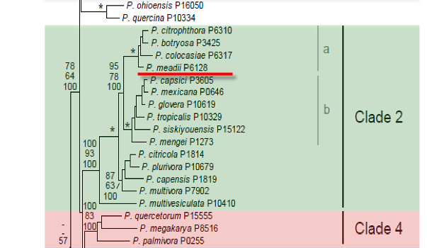
Genus wide phylogeny for Phytophthora using four mitochondrial loci (cox2, nad9, rps10 and secY; 2,373 nucleotides). Maximum likelihood branch lengths shown. Numbers on nodes represent bootstrap support values for maximum likelihood (top), maximum parsimony (middle) and Bayesian posterior probabilities as percentages (bottom). Nodes receiving significant support (>95%) in all analysis are marked with an asterisk (*). Scale bar indicates number of substitutions per site.(Martin, Blair and Coffey, unpublished).

[ Click the tree to enlarge it. ]
Phytophthora meadii McRae 1918 (Oomycetes, Pythiales)
Notes: Listed by Tucker (1931) as a synonym of Phytophthora palmivora, but considered by Waterhouse (1963) to be a distinct species.
Distribution: Asia, Australia, Pacific Islands (USA: HI).
Substrate: Fruits (pods), stems, inflorescences.
Disease Note: Abnormal leaf fall of rubber.
Host: Various plant families including Hevea spp. (rubber, Euphorbiaceae).
Supporting Literature:
Stamps, D.J. 1985. Phytophthora meadii. C.M.I. Descript. Pathog. Fungi Bact. 834: 1-2
Tucker, C.M. 1931. Taxonomy of the genus Phytophthora de Bary. Univ. Missouri Agric. Exp. Sta. Bull. 153: 1-208
Waterhouse, G.M. 1963. Key to the species of Phytophthora de Bary. Mycol. Pap. 92: 1-22
Updated on May 26, 2006
P. meadii is classified in group II (Stamps et al. 1990). A summary of characteristics is given by Waterhouse (1974b), Stamps (1985d), and Holliday (1980). Morphology is shown in Figure 1. See Tables 4.2 and 4.3 for tabular keys, Appendix 4.9 for a dichotomous key (Ho 1992), and Table 1 and Table 2 for morphological comparison with P. palmivora and related species in Phytophthora Diseases Worldwide (Erwin and Ribeiro 1996). Sansome et al. (1991) present a hypothesis that P. meadii might be a species hybrid with P. palmivora as one parent because self-fertile isolates of P. meadii were polyploid but A1 and A2 types were diploid; however, Oudemans and Coffey (1991b) indicated by isozyme data that P. palmivora would be an unlikely parent because of the large genetic difference separating these species.
1. Sporangia
Sporangia are terminal or lateral; papillate, occasionally with two papillae; caducous (pedicel is 10 to 20 µm in length); ellipsoid or elongated, obpyriform, occasionally spherical, often distorted into lobed or hourglass shapes; on fruit they are 33 to 67 µm long x 14 to 28 µm wide (average 48 x 21 µm) and in water 20 to 44 µm long x 16 to 29 µm wide (average 32 x 23 µm). The length-breadth ratio varies from 1.3 to 2.0 (Waterhouse 1974a). Oudemans and Coffey (1991b) report a range of 1.4 to 1.7. Sporangia are produced sympodially on branched sporangiophores that are 10 to 20 µm long (Figure 1).
2. Hyphal Swellings
Hyphal swellings are not produced.
3. Chlamydospores
Chlamydospores are rare and range from 16 to 30 µm in diameter with the majority under 30 µm. They are usually produced at temperatures above 30oC after periods of incubation >18 days (Peries and Fernando 1966).
4. Sex Organs
P. meadii is predominantly homothallic (Waterhouse 1974b) with some evidence of heterothallism (Peries and Dantanarayana 1965). A1 and A2 types have been described in India (Rajalakshmy et al. 1985). A1 isolates of P. meadii paired with A2 isolates of P. palmivora produced oospores (Sastry and Hegde 1987c). Sex organs developed only at 15 to 20oC (Peries and Fernando 1966). Antheridia are rounded and amphigynous (12 x 13 up to 10 x 16 µm in diameter); oogonia are spherical to pyriform with smooth or wrinkled walls; those produced in agar culture measure 21 to 49 µm in diameter (average 33.4 µm); on fruit, oogonia measure 20 to 48 µm in diameter (average 25.1 µm); oospores are markedly aplerotic; those produced in agar media measure 16 to 32.8 µm in diameter (average 24 µm); on fruit, oospores measure 16 to 32 µm in diameter (average 21 µm), and the oospore wall is 4 µm thick.
5. Growth Temperatures
The minimum temperature for growth is >5oC, the optimum is 25 to 30oC, and the maximum is >33oC.
6. Distinguishing Characteristics
Dantanarayana et al. (1984) distinguished P. meadii from P. palmivora by production of spherical to ovoid, caducous sporangia on medium-length pedicels (P. palmivora sporangia have short pedicels; Figure 4.15 in Phytophthora Diseases Worldwide [Erwin and Ribeiro 1996]), rare production of chlamydospores, absence of pigment production on agar media, formation of the sympodium on which sporangia formed, and formation of aplerotic oospores (Table 1). Prior to Waterhouse (1963, 1974a) and Dantanarayana et al. (1984), many isolations from rubber in Sri Lanka and southern India were erroneously ascribed to P. palmivora because P. meadii was not considered by some to be distinct (Holliday 1980).
Oudemans and Coffey (1991b) reported that sporangia of P. meadii (six isolates) were slightly larger (average 38.5 x 24.6 µm) than those of P. botryosa (four isolates; average 29.8 x 17.9 µm); however, isozyme data of Oudemans and Coffey (1991b) indicate that P. meadii and P. botryosa have a high level of similarity but that P. meadii is genetically distinct from P. palmivora. They suggest that P. meadii and P. botryosa probably represent two geographically separate populations of a single species, since P. meadii has been reported mainly from India and Sri Lanka and P. botryosa from Thailand, Malaysia, and Vietnam. See Figure 4.15 in Phytophthora Diseases Worldwide (Erwin and Ribeiro 1996), in which the sporangial morphology of these three species is compared. Table 1 and Table 2 (Erwin and Ribeiro 1996) give morphological data for P. meadii and several other similar species.
Nomenclature information was provided by the the Systematic Botany and Mycology Laboratory in USDA-ARS.
Isolate list