
Phytophthora has been rebuilt to fix security-related problems and to restore GIS tools. These tools allow users to visualize the geospatial, temporal, and environmental contexts of Phytophthora discoveries. The next phase is to update species information and add data derived from large-scale surveys. If you have suggestions and requests to make the database better, please contact Seogchan Kang (sxk55@psu.edu).
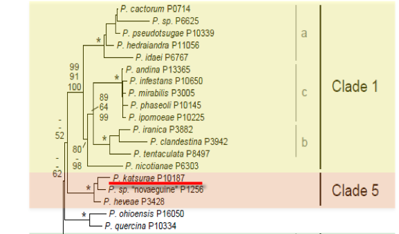
Genus wide phylogeny for Phytophthora using four mitochondrial loci (cox2, nad9, rps10 and secY; 2,373 nucleotides). Maximum likelihood branch lengths shown. Numbers on nodes represent bootstrap support values for maximum likelihood (top), maximum parsimony (middle) and Bayesian posterior probabilities as percentages (bottom). Nodes receiving significant support (>95%) in all analysis are marked with an asterisk (*). Scale bar indicates number of substitutions per site.(Martin, Blair and Coffey, unpublished).

[ Click the tree to enlarge it. ]
Phytophthora katsurae W.H. Ko & H.S. Chang 1979 (Oomycetes, Pythiales)
[ Phytophthora castaneae Katsura & K. Uchida 1968 - illegitimate later homonym, not included in search] Note: Illegitimate later homonym of Phytophthora castaneae (Mangis) Clements & Shear 1931.
Notes: Phytophthora katsurae was published as a replacement name when it was discovered that Phytophthora castaneae Katsura & K. Uchida was an illegitimate later homonym of Phytophthora castaneae (Mangis) Clements & Shear 1931.
Distribution: Africa (Cote d\\'Ivoire), Asia (Japan, Taiwan), Caribbean (Jamaica), Pacific Islands (USA: HI), Papua New Guinea.
Substrate: Tree trunks, pods, fruit.
Disease Note: Trunk rot of chestnut, fruit and heart rot of coconut.
Host: Cocos nucifera (coconut, Arecaceae); Castanea crenata (chestnut, Fagaceae). Also reported from Theobroma cacao (cacao, Malvaceae) (Erwin & Ribeiro 1996).
Supporting Literature:
Erwin, D.C., and Ribeiro, O.K. 1996. Phytophthora Diseases Worldwide. APS Press, St. Paul, Minnesota, 562 pages.
Kroon, L.P.N.M., Bakker, F.T., van den Bosch, G.B.M., Bonants, P.J.M., and Flier, W.G. 2004. Phylogenetic analysis of Phytophthora species based on mitochondrial and nuclear DNS sequences. Fungal Genet. Biol. 41: 766-782
Stamps, D.J. 1985. Phytophthora katsurea. C.M.I. Descript. Pathog. Fungi Bact. 837: 1-2
Waterhouse, G.M. 1963. Key to the species of Phytophthora de Bary. Mycol. Pap. 92: 1-22
Updated on Jun 09, 2006
P. katsurae is classified in group II (Stamps et al. 1990). See Tables 4.2 and 4.3 for tabular keys and Appendix 4.9 for a dichotomous key (Ho 1992) in Phytophthora Diseases Worldwide (Erwin and Ribeiro 1996). Morphology is shown in Figure 1 and Figure 2.
1. Sporangia
Sporangia are mostly terminal but occasionally are laterally attached and sometimes formed intercalarily. They are limoniform, ovoid, obpyriform to obturbinate, and papillate, occasionally with two papillae per sporangium. Some sporangia are asymmetric (Figures 39.1 and 39.2); they are 10 to 42.5 µm long x 10 to 37.5 µm wide (average 27.5 x 22.5 µm) on the stalk (Katsura 1976). Ko and Chang (1979) recorded sizes of sporangia as 25.1 to 47.4 x 22.3 to 30.7 µm. Length-breadth ratio is <2.0. Sporangiophores are unbranched.
2. Hyphal Swellings
Hyphal swellings are not formed.
3. Chlamydospores
Chlamydospores are spherical and 12.0 to 19.2 µm in diameter.
4. Sex Organs
P. katsurae is homothallic. Oospores form abundantly in 10% V8 juice agar (Ko and Chang 1979). Antheridia are amphigynous and spherical to ovoid; oogonia have warty protuberances on the surface and a distinctf unnel-shaped base (Figures 1 and 2); oogonia are 19 to 31 µm in diameter (average 27 x 25 µm); oospores are globose, 15 to 27.5 µm in diameter (average 20 µm). Up to 95% of 30-day-old oospores germinated when incubated under blue and green light (H. S. Chang and Shu 1988).
5. Growth Temperatures
The minimum temperature for growth is 9oC, optimum 26 to 28oC, and maximum 32oC.
6. Distinguishing Characteristics
P. katsurae differs from P. cambivora, which is also a pathogen of chestnut trees (see Chapter 13 in Phytophthora Diseases Worldwide [Erwin and Ribeiro 1996]), in that it is homothallic and the oogonia and oospores are smaller. Sporangia of P. katsurae are papillate, whereas those of P. cambivora are nonpapillate. It differs from P. heveae by the presence of protuberances on the oogonial wall and the funnel-shaped oogonial stalk (Katsura 1976; also noted by Oudemans and Coffey [1991b]). See Chapter 4, Figure 4.28 in Phytophthora Diseases Worldwide (Erwin and Ribeiro 1996), for a photomicrograph comparing oogonia of P. heveae and P. katsurae (Oudemans and Coffey 1991b). Oudemans and Coffey (1991b) found by isozyme analysis that P. heveae and P. katsurae are closely related.
Nomenclature information was provided by the the Systematic Botany and Mycology Laboratory in USDA-ARS.
Isolate list