
Phytophthora has been rebuilt to fix security-related problems and to restore GIS tools. These tools allow users to visualize the geospatial, temporal, and environmental contexts of Phytophthora discoveries. The next phase is to update species information and add data derived from large-scale surveys. If you have suggestions and requests to make the database better, please contact Seogchan Kang (sxk55@psu.edu).
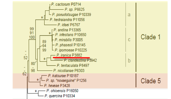
Genus wide phylogeny for Phytophthora using four mitochondrial loci (cox2, nad9, rps10 and secY; 2,373 nucleotides). Maximum likelihood branch lengths shown. Numbers on nodes represent bootstrap support values for maximum likelihood (top), maximum parsimony (middle) and Bayesian posterior probabilities as percentages (bottom). Nodes receiving significant support (>95%) in all analysis are marked with an asterisk (*). Scale bar indicates number of substitutions per site.(Martin, Blair and Coffey, unpublished).

[ Click the tree to enlarge it. ]
Phytophthora iranica Ershad 1971 (Oomycetes, Pythiales)
Notes: Isolates from Myrtus communis in Italy were initially reported as Phytophthora iranica, but are now considered to be a distinct species, Phytophthora italica.
Distribution: Asia (Iran). Reports from Italy are the new species Phytophthora italica.
Substrate: Roots, fruits, tubers.
Disease Note: Root and stem rot, tuber soft rot.
Host: Solanum melongena (eggplant); Solanum tuberosum (potato), Lycopersicon esculentum (tomato), all Solanaceae, and Beta vulgaris (sugar beet, Chenopodiaceae) are infected when inoculated. Reports on Myrtus (Myrtaceae) are Phytophthora italica.
Supporting Literature:
Erwin, D.C., and Ribeiro, O.K. 1996. Phytophthora Diseases Worldwide. APS Press, St. Paul, Minnesota, 562 pages.
Kroon, L.P.N.M., Bakker, F.T., van den Bosch, G.B.M., Bonants, P.J.M., and Flier, W.G. 2004. Phylogenetic analysis of Phytophthora species based on mitochondrial and nuclear DNS sequences. Fungal Genet. Biol. 41: 766-782
Updated on Jun 14, 2006
P. iranica is classified in group I (Stamps et al. 1990). Morphology is shown in Figure 1 for an isolate from Iran. See Tables 4.2 and 4.3 in Phytophthora Diseases Worldwide (Erwin and Ribeiro 1996) for tabular keys.
1. Sporangia
Sporangia are papillate (sometimes with two papillae per sporangium); ovoid, obpyriform, ellipsoid to subspherical, 30 to 72 µm long x 22 to 51 µm wide (average 47.9 x 36.8 µm) (length-breadth ratio 1.3:1); and persistent on the stalk. Sporangiophores are short and sympodially branched (Ershad 1971).
2. Chlamydospores
Chlamydospores are rare; 17 to 41 µm in diameter, average 28.7 µm; mostly intercalary; and rarely terminal.
3. Hyphal Swellings
Hyphal swellings are not formed.
4. Sex Organs
Oogonia are formed in single culture (homothallic) and are subspherical and 21 to 45 µm in diameter, average 34 µm. Antheridia are mostly paragynous, but occasionally an amphigynous antheridium is found; oospores are aplerotic, 15 to 37 µm in diameter, average 29.3 µm; the oospore wall is 1 to 5 µm thick, average 3 µm (Ershad 1971).
5. Growth Temperatures
The minimum temperature for growth is 10oC, optimum 27.5oC, and maximum 35oC (Ershad 1971). Similar values were reported by Belisario et al. (1993).
6. Distinguishing Characteristics
P. iranica differs from P. cactorum by its production of noncaducuous sporangia, sporangia with more than one papilla, and thicker oospore walls.
When tested by electrophoresis on slabs of polyacrylamide gel, P. iranica (now P. italica) produced protein patterns clearly distinguishable from those of P. citrophthora, P. cactorum, and P. nicotianae (Belisario et al. 1993).
Nomenclature information was provided by the the Systematic Botany and Mycology Laboratory in USDA-ARS.
Isolate list