
Phytophthora has been rebuilt to fix security-related problems and to restore GIS tools. These tools allow users to visualize the geospatial, temporal, and environmental contexts of Phytophthora discoveries. The next phase is to update species information and add data derived from large-scale surveys. If you have suggestions and requests to make the database better, please contact Seogchan Kang (sxk55@psu.edu).
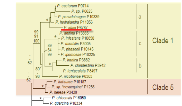
Genus wide phylogeny for Phytophthora using four mitochondrial loci (cox2, nad9, rps10 and secY; 2,373 nucleotides). Maximum likelihood branch lengths shown. Numbers on nodes represent bootstrap support values for maximum likelihood (top), maximum parsimony (middle) and Bayesian posterior probabilities as percentages (bottom). Nodes receiving significant support (>95%) in all analysis are marked with an asterisk (*). Scale bar indicates number of substitutions per site.(Martin, Blair and Coffey, unpublished).

[ Click the tree to enlarge it. ]
Phytophthora idaei D.M. Kenn. 1995 (Oomycetes, Pythiales)
Distribution: Europe (UK).
Substrate: Roots.
Disease Note: Root rot. No aerial symptoms were produced when up to 20 percent of roots were infected; potentially spread by cultivation of apparently healthy plants.
Host: Rubus idaeus (red raspberry, Rosaceae).
Supporting Literature:
Erwin, D.C., and Ribeiro, O.K. 1996. Phytophthora Diseases Worldwide. APS Press, St. Paul, Minnesota, 562 pages.
Kroon, L.P.N.M., Bakker, F.T., van den Bosch, G.B.M., Bonants, P.J.M., and Flier, W.G. 2004. Phylogenetic analysis of Phytophthora species based on mitochondrial and nuclear DNS sequences. Fungal Genet. Biol. 41: 766-782
Updated on Jun 13, 2006
Kennedy and Duncan (1995) classified P. idaei in group I (Stamps et al. 1990). A comparison of the morphology of sporangia, sporangiophore branching, and sexual organs of P. idaei, P. cactorum, P. citricola, and P. syringae is shown in Figure 1 (Kennedy and Duncan 1995). See Tables 4.2 and 4.3 for tabular keys in Phytophthora Diseases Worldwide (Erwin and Ribeiro 1996). Colonies of P. idaei have limited aerial mycelium. Chlamydospores and hyphal swellings are not formed.
1. Sporangia
Sporangia form optimally at 20°C and are papillate and predominantly spherical to ovoid (49 x 36 µm). Germination by production of zoospores is most common. Direct germination is rare. Sporangiophores are simple and in a lax sympodium. Sporangia are persistent (noncaducous).
2. Sex Organs
Oogonia (26 x 38 µm) form in single culture (homothallic) and in the cortex of roots of infected raspberry plants and are smooth and spherical. Oospores (22-34 µm) are aplerotic in the oogonia. Antheridia are predominantly paragynous.
3. Growth Temperatures
Growth, sporangial formation, and oogonial and oospore formation are optimal at 20-22°C.
4. Distinguishing Characteristics
P. idaei resembles P. cactorum because the sporangia are nearly spherical and are papillate. Also, antheridia of both species are paragynous. Sporangia of P. idaei are persistent while those of P. cactorum are caducous. Hyphae of P. idaei are consistently wider than those of P. cactorum. The optimal temperature for sporulation and growth is lower for P. idaei (about 20°C) than for P. cactorum (about 25-28°C). P. idaei isolates do not utilize nitrate as a nitrogen source, whereas P. cactorum isolates do.
P. idaei differs from P. iranica by its failure to produce chlamydospores and from P. pseudotsugae by its much lower production of sporangia in liquid culture. P. idaei differs from P. clandestina by its absence of hyphal swellings, persistent sporangia, subterminal or digitate antheridia, and much lower levels of amphigyny (see also Taylor et al. [1983] for a description of P. clandestina).
Nomenclature information was provided by the the Systematic Botany and Mycology Laboratory in USDA-ARS.
Isolate list