
Phytophthora has been rebuilt to fix security-related problems and to restore GIS tools. These tools allow users to visualize the geospatial, temporal, and environmental contexts of Phytophthora discoveries. The next phase is to update species information and add data derived from large-scale surveys. If you have suggestions and requests to make the database better, please contact Seogchan Kang (sxk55@psu.edu).
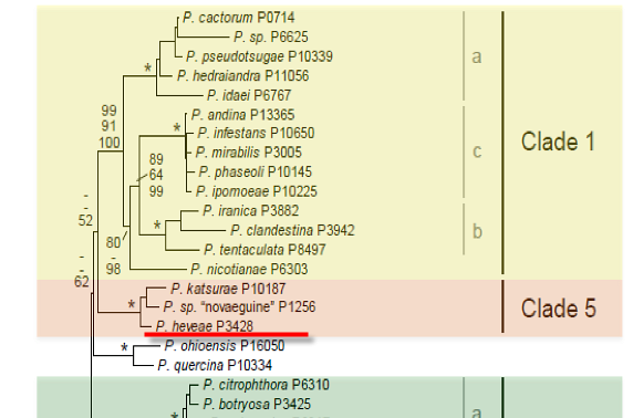
Genus wide phylogeny for Phytophthora using four mitochondrial loci (cox2, nad9, rps10 and secY; 2,373 nucleotides). Maximum likelihood branch lengths shown. Numbers on nodes represent bootstrap support values for maximum likelihood (top), maximum parsimony (middle) and Bayesian posterior probabilities as percentages (bottom). Nodes receiving significant support (>95%) in all analysis are marked with an asterisk (*). Scale bar indicates number of substitutions per site.(Martin, Blair and Coffey, unpublished).

[ Click the tree to enlarge it. ]
Phytophthora heveae A. Thomps. 1929 (Oomycetes, Pythiales) Phytophthora palmivora var. heveae (A.W. Thomps.) Orellana 1959 Note: See Waterhouse 1963.
Distribution: Asia Australiasia North America (USA: NC), South America (Brazil).
Substrate: Roots, leaves, buds, seeds, pods, stems.
Disease Note: Rot, leaf blight and stem canker. Disease symptoms on crop species include black stripe of rubber bud rot and nut fall of coconut and dieback of rhododendron (Erwin & Ribeiro 1996).
Host: 12 genera in 11 families including Hevea (rubber Euphorbiaceae) Rhododendron (Ericaceae) and Cocos nucifera (coconut Arecaceae).
Supporting Literature:
Erwin, D.C., and Ribeiro, O.K. 1996. Phytophthora Diseases Worldwide. APS Press, St. Paul, Minnesota, 562 pages.
Kroon, L.P.N.M., Bakker, F.T., van den Bosch, G.B.M., Bonants, P.J.M., and Flier, W.G. 2004. Phylogenetic analysis of Phytophthora species based on mitochondrial and nuclear DNS sequences. Fungal Genet. Biol. 41: 766-782
Stamps, D.J. 1978. Phytophthora heveae. C.M.I. Descript. Pathog. Fungi Bact. 594: 1-2
Waterhouse, G.M. 1963. Key to the species of Phytophthora de Bary. Mycol. Pap. 92: 1-22
Updated on Jun 07, 2006
P. heveae is classified in group II (Stamps et al. 1990), and its characteristics are summarized by Waterhouse (1974c) and Stamps (1978d). Morphology is illustrated in Figure 1. See Tables 4.2 and 4.for tabular keys and Appendix 4.9 for a dichotomous key (Ho 1992) in Phytophthora Diseases Worldwide (Erwin and Ribeiro 1996). Its characteristics are compared with P. palmivora and several other species of Phytophthora in Tables 51A.4 and 51A.5 in Phytophthora Diseases Worldwide (Erwin and Ribeiro 1996). Zentmyer et al. (1978) discuss the characteristics of several isolates. Zhang et al. (1995) summarize the characteristics of P. heveae as reported by several workers.
1. Sporangia
Sporangia are papillate, irregular in shape, obpyriform to ellipsoidal, and often asymmetrical and form laterally on the stalk. They are 27 to 66 µm long x 20 to 49 µm wide (average 45 x 29.6 µm) with a length-breadth ratio of 1.1 to 2.9:1 (average 1.5) and caducous (pedicel <10 µm) (Albuquerque et al. 1974). However, in isolates obtained from avocado as well as in several reference isolates, caducity was not noted, and sporangiophores that did detach from the stalk were of varying length (Zentmyer et al. 1978). Caducity was not noted by Thompson (1929). Sporangia develop profusely on agar media. Light increased sporangium production in vitro (Brasier 1969a). Branching of sporangiophores is irregular, and intercalary swellings are often present.
2. Chlamydospores
Chlamydospores are not produced.
3. Sex Organs
Hyphal swellings are small to large, resembling abortive reproductive organs. P. heveae is homothallic. Antheridia are amphigynous, usually spherical, occasionally cylindrical (11 x 10 µm), and sometimes bicellular (reported first by Zentmyer et al. [1978] on an avocado isolate and on authentic reference isolates). Although Albuquerque et al. (1974) noted some paragynous antheridia, none were seen by Zentmyer et al. (1978) in eight cultures from rubber, cocoa, rhododendron, and avocado. Oogonia are globose and form readily in culture, often in close groups; they are broadly funnel shaped at the base and 17 to 32 µm in diameter (average 22.3 µm) (see Figure 4.285 in Erwin and Ribeiro (1996, Phytophthora Diseases Worldwide).). Oospores are round, smooth, thick walled, 15 to 26.8 µm in diameter (average 21.5 µm), and markedly aplerotic (see Stamps 1978d and Holliday 1980). Zentmyer et al. (1978) noted that the average oospore diameter of isolate P640 from rubber on carrot agar was 23 µm, smaller than the 28-µm average diameter cited by Waterhouse (1963), and that this difference was probably the result of the use of carrot medium rather than cornmeal. Berg et al. (1966) and Leal et al. (1965) described oospore germination. Up to 90% of 30-day-old oospores germinated when they were incubated under blue and green light (Chang and Shea 1988).
4. Growth Temperatures
The minimum temperature for growth is 11.5oC, optimum 25oC, and maximum 31.0 to 32.5oC. Glutamine, urea, and ethanol reduced growth in synthetic media (Leal et al. 1968).
5. Distinguishing Characteristics
P. heveae is distinguished from P. meadii by lack of aerial mycelium, funnel-shaped oogonia, smaller and more ratio of sporangia is greater in P. heveae). P. heveae is distinguished from P. colocasiae, P. parasitica, and P. palmivora by the lack of aerial mycelium and by the absence of chlamydospores (see Table 51A.4 in Erwin and Ribeiro (1996, Phytophthora Diseases Worldwide) for a comparison of characteristics with other species).
Nomenclature information was provided by the the Systematic Botany and Mycology Laboratory in USDA-ARS.
Isolate list