
Phytophthora has been rebuilt to fix security-related problems and to restore GIS tools. These tools allow users to visualize the geospatial, temporal, and environmental contexts of Phytophthora discoveries. The next phase is to update species information and add data derived from large-scale surveys. If you have suggestions and requests to make the database better, please contact Seogchan Kang (sxk55@psu.edu).
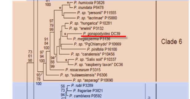
Genus wide phylogeny for Phytophthora using four mitochondrial loci (cox2, nad9, rps10 and secY; 2,373 nucleotides). Maximum likelihood branch lengths shown. Numbers on nodes represent bootstrap support values for maximum likelihood (top), maximum parsimony (middle) and Bayesian posterior probabilities as percentages (bottom). Nodes receiving significant support (>95%) in all analysis are marked with an asterisk (*). Scale bar indicates number of substitutions per site.(Martin, Blair and Coffey, unpublished).

[ Click the tree to enlarge it. ]
Phytophthora gonapodyides (H.E. Petersen) Buisman 1927 (Oomycetes, Pythiales) Pythiomorpha gonapodyides H.E. Petersen 1909
Variant spelling Pythiomorpha gonapodyoides H.E. Petersen 1909
Notes: Possibly reported on Pinaceae seedlings under the name Phytophthora drechsleri (Erwin & Ribeiro 1996).
Distribution: North America (USA), Europe (Denmark). Also reported from Australia, New Zealand, and South America (Chile).
Substrate: Roots, seedlings.
Disease Note: Root rot. A minor pathogen (Erwin & Ribeiro 1996).
Host: Thirteen genera in 11 families, including Malus spp. (Rosaceae); possibly also on Pinaceae seedlings including Pseudotsuga menziesii (Douglas-fir), Abies spp., and Tsuga mertensiana (mountain hemlock).
Supporting Literature:
Erwin, D.C., and Ribeiro, O.K. 1996. Phytophthora Diseases Worldwide. APS Press, St. Paul, Minnesota, 562 pages.
Kroon, L.P.N.M., Bakker, F.T., van den Bosch, G.B.M., Bonants, P.J.M., and Flier, W.G. 2004. Phylogenetic analysis of Phytophthora species based on mitochondrial and nuclear DNS sequences. Fungal Genet. Biol. 41: 766-782
Waterhouse, G.M. 1970. The genus Phytophthora de Bary. Mycol. Pap. 122: 1-59
Updated on Jun 12, 2006
P. gonapodyides is classified in group VI (Stamps et al. 1990). See Tables 4.2 and 4.3 in Phytophthora Diseases Worldwide (Erwin and Ribeiro 1996) for tabular keys. Morphology of spores is shown in Figure 1 and Figure 2A-C. A typical colony is shown in Figure 2D. See Waterhouse (1958) for a comprehensive description.
1. Sporangia
Sporangia are nonpapillate, proliferating internally; nondeciduous; ellipsoid, ovoid, to obpyriform; and 42 to 75 µm long x 20 to 32 µm wide (average 44 x 22 µm) (Petersen 1910). Sporangia are broadly pyriform, elongated pyriform, and occasionally ovate, 23 to 48 µm x 43 to 91 µm, mean 35.8 x 58.7 µm. The length-breadth ratio is 1.2:1.0 to 2.4:1.0, mean 1.6:1.0. Proliferation may be internal, nested, or external. Sporangiophores are sympodial only in water (Pittis and Colhoun 1984).
2. Chlamydospores
Niether chlamydospores or hyphal swellings are produced.
3. Sex Organs
P. gonapodyides is heterothallic; however, Kanouse (1925) reported that oogonia were produced in single culture, but this report has not been confirmed (Waterhouse 1958). Oogonia with amphigynous antheridia were produced by pairing an A1 culture with an A2 culture of P. drechsleri. Oogonia are globose (27 to 48 µm in diameter, mean 40.1 µm). Walls are frequently rough (Figure 2C). Antheridia are amphigynous and variable in shape. Oospores in culture are aplerotic, thick walled, 18 to 32 µm in diameter, mean 21.1 µm, and yellow (Pittis and Colhoun 1984). All isolates studied by Brasier et al. (1993) were self sterile but when paired with A2 isolates of P. cambivora, P. megakarya, or P. meadii produced gametangia that were characteristic of these species.
4. Growth Temperatures
Mycelial growth forms a rosette pattern (Figure 2D). The minimum temperature for growth is <5oC, optimum 25oC, and maximum 30 to 35oC. Brasier et al. (1993) reported slow growth at 20, 25, and 30oC but no growth at 35oC.
5. Distinguishing characteristics
[[PAPER:2352|1]]
Nomenclature information was provided by the the Systematic Botany and Mycology Laboratory in USDA-ARS.
Isolate list