
Phytophthora has been rebuilt to fix security-related problems and to restore GIS tools. These tools allow users to visualize the geospatial, temporal, and environmental contexts of Phytophthora discoveries. The next phase is to update species information and add data derived from large-scale surveys. If you have suggestions and requests to make the database better, please contact Seogchan Kang (sxk55@psu.edu).
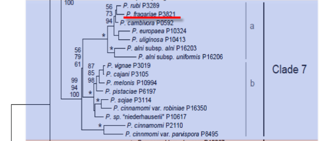
Genus wide phylogeny for Phytophthora using four mitochondrial loci (cox2, nad9, rps10 and secY; 2,373 nucleotides). Maximum likelihood branch lengths shown. Numbers on nodes represent bootstrap support values for maximum likelihood (top), maximum parsimony (middle) and Bayesian posterior probabilities as percentages (bottom). Nodes receiving significant support (>95%) in all analysis are marked with an asterisk (*). Scale bar indicates number of substitutions per site.(Martin, Blair and Coffey, unpublished).

[ Click the tree to enlarge it. ]
Phytophthora fragariae var. fragariae Hickman 1940 (Oomycetes, Pythiales)
=Phytophthora fragariae C.J. Hickman 1940 Note: See type variety.
Distribution: Asia, Australia, New Zealand, Europe, North America (Canada, USA).
Substrate: Roots.
Disease Note: Red stele or red core root rot. The most important fungal pathogen of strawberries (Ho & Jong 1988). Foreign races are listed by APHIS as a regulated plant pest.
Host: Fragaria x ananassa and Rubus ursinus var. longanobaccus (Rosaceae) are the only known hosts under natural conditions. Infection of other Rosaceae, Chenopodiaceae, and Solanaceae can occur via artificial inoculation (Erwin 1996).
Supporting Literature:
Erwin, D.C., and Ribeiro, O.K. 1996. Phytophthora Diseases Worldwide. APS Press, St. Paul, Minnesota, 562 pages.
Ho, H.H., and Jong, S.C. 1988. Phytophthora fragariae. Mycotaxon 31: 305-322
Updated on Oct 13, 2005
P. fragariae var. rubi is classified in group V (Stamps et al. 1990). See Tables 4.2 and 4.3 in Phytophthora Diseases Worldwide (Erwin and Ribeiro 1996) for tabular keys.
Duncan et al. (1991) showed that P. fragariae var. rubi is similar to P. fragariae in production of sporangia and oospores and in temperature relations. Electrophoretic assay on polyacrylamide gels showed only slight differences in protein patterns. Although P. fragariae var. rubi was distinguishable from P. fragariae var. fragariae by DNA technology, most mtDNA and nuclear DNA-developed probes from both varieties hybridized, thus indicating that they are related. That some hybridization occurred with P. cambivora indicated that its relationship with P. fragariae should be investigated further (Stammler et al. 1993).
A comparative study of the molecular characterization by DNA restriction fragment length polymorphisms (RFLP) of the strawberry and red raspberry biotypes showed a 54 to 55% similarity between the two groups but did not show a close genetic relationship with P. megasperma or P. erythroseptica, which had previously been named as causal agents (FÖrster and Coffey 1992).
In an analysis of a wide range of P. fragariae var. fragariae (from strawberry) and P. fragariae var. rubi isolates via Southern blot hybridization, DNA from P. fragariae var. fragariae, which had been cloned in Escherichia coli bacteria, hybridized with P. fragariae var. rubi; however, little or no hybridization occurred with DNA of other Phytophthora species, including P. megasperma and P. erythroseptica (Stammler et al. 1993). Since isolates of either P. fragariae var. fragariae or P. fragariae var. rubi were very homogenous (little variability), it could be assumed that both populations had been spread worldwide from a common source, probably by horticultural movement of plant material. The 54-55% similarity in mtDNA RFLP patterns of P. fragariae from strawberry and P. fragariae var. rubi from red raspberry supports the hypothesis by Duncan et al. (1991) that both groups might have evolved from a common ancestor pathogenic on wild members of the tribe Potentilleae in the Pacific Northwest of the United States (FÖrster and Coffey 1992). Wilhelm and Sagen (1974) suggested that the red stele disease of strawberry caused by P. fragariae probably had its origin in this area.
The description of the new variety, P. fragariae var. rubi, is given below (reproduced by permission of the British Mycological Society, Manchester, United Kingdom, courtesy of Wilcox et al. 1993):
1. Hyphae
Hyphae hyaline, non-septate or sparingly so in older parts: width 5.3 µm to 7.4 µm, mean 6.5 µm.
2. Sporangia
Sporangiophores simple or sparingly branched. Sporangia produced only when irrigated; non-deciduous, terminal, non-papillate, proliferating internally by nesting and extension, and less often in sympodial fashion on a short lateral branch immediately beneath the original sporangium; ovoid to obpyriform in shape; sometimes tapered at the base; usually 53-68 x 34-44 µm (59 x 40 µm); wall thin with slight apical thickening; germinating by zoospores, rarely by germ tube. Zoospores 13 x 10 µm.
3. Sexual organs
Sexual organs formed in abundance in the vascular tissue of raspberry roots; also formed on some agar media, sometimes more commonly in the area around the inoculum plug. Oogonial terminal, usually globose with a tapered base 30-45 µm in diam. (mean ca. 39 µm); becoming golden brown with age. Antheridia terminal, amphigynous or less commonly paragynous; sphaeroidal to ovoidal; averaging 13-16 µm.Oospores spherical and aplerotic; 28-37 µm diam. (mean ca. 35 µm) with a smooth hyaline wall 2.7-3.4 µm (mean 3.0).
4. Growth temperatures
Minimum growth at or below 4°C, optimum 19-22°C, and maximum 25-28°C.
5. Growth characteristics in culture media
Mycelial colonies grow slowly but well on sucrose asparagine, french bean, lima bean, and V8-juice agars, but growth is poor or absent on media with nitrate as the sole nitrogen source. Colonies uniform with profuse aerial mycelium on most agars including sucrose asparagine and V8 agars.
6. Distinguishing characteristics
The habitat of P. fragariae var. rubi is in the roots, crown, and canes of cultivated raspberries in most countries of northern Europe and North America. The varietal epithet of this fungus is named for its host, Rubus spp. The primary cultural traits distinguishing P. fragariae var. rubi from P. fragariae var. fragariae are the former\'s more robust growth on agar media and far greater tendency to form oospores in single agar culture. The two varieties also differ in the electrophoretic patterns of their mycelial proteins, in the composition of their mitochondrial and nuclear DNA as indicated by RFLP analysis and hybridization studies, and in host range, with Rubus and Fragaria spp. as primary hosts, respectively.
[[PAPER:2351|1]]
[[PAPER:2301|1]]
Nomenclature information was provided by the the Systematic Botany and Mycology Laboratory in USDA-ARS.
Isolate list