
Phytophthora has been rebuilt to fix security-related problems and to restore GIS tools. These tools allow users to visualize the geospatial, temporal, and environmental contexts of Phytophthora discoveries. The next phase is to update species information and add data derived from large-scale surveys. If you have suggestions and requests to make the database better, please contact Seogchan Kang (sxk55@psu.edu).
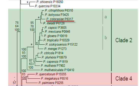
Genus wide phylogeny for Phytophthora using four mitochondrial loci (cox2, nad9, rps10 and secY; 2,373 nucleotides). Maximum likelihood branch lengths shown. Numbers on nodes represent bootstrap support values for maximum likelihood (top), maximum parsimony (middle) and Bayesian posterior probabilities as percentages (bottom). Nodes receiving significant support (>95%) in all analysis are marked with an asterisk (*). Scale bar indicates number of substitutions per site.(Martin, Blair and Coffey, unpublished).

[ Click the tree to enlarge it. ]
Phytophthora colocasiae Racib. 1900 (Oomycetes, Pythiales) Kawakamia colocasiae (Racib.) Sawada 1911Phytophthora parasitica var. colocasiae (Racib.) Sarej. 1936
Distribution: Africa (Ethiopia, East Africa, Fernando Po), Asia, Caribbean (Dominican Republic), North America (USA: CA, NC, HI), Pacific Islands, South America (Brazil, Argentina).
Substrate: Leaves, corms, stems.
Disease Note: Leaf blight, corm rot, wilt; also stem canker and black stripe of rubber.
Host: Colocasia esculenta (taro) and other Aracaceae; also six genera in five other families.
Supporting Literature:
Erwin, D.C., and Ribeiro, O.K. 1996. Phytophthora Diseases Worldwide. APS Press, St. Paul, Minnesota, 562 pages.
Ho, H.H., and Chang, H.S. 1992. A re-evaluation of Phytophthora species described by K. Sawada in Taiwan. Mycotaxon 43: 297-316
Kroon, L.P.N.M., Bakker, F.T., van den Bosch, G.B.M., Bonants, P.J.M., and Flier, W.G. 2004. Phylogenetic analysis of Phytophthora species based on mitochondrial and nuclear DNS sequences. Fungal Genet. Biol. 41: 766-782
Updated on Jun 12, 2006
P. colocasiae is classified in group IV (Stamps et al. 1990). See Tables 4.2 and 4.3 for tabular keys and Appendix 4.9 for a dichotomous key (Ho 1992) in Phytophthora Diseases Worldwide (Erwin and Ribeiro 1996). Morphology is shown in Figure 1.
1. Sporangia
Sporangia are semipapillate; caducous (pedicel length 3.5 to 10 µm); ovoid, ellipsoid, or sometimes fusiform; and 40 to 70 µm long x 17 to 28 µm wide. Sawada (1911) reported sporangia 114 µm in length, average 59 x 23 µm. They have a length-breadth ratio of 1.6:1 and a tapered base with an occasional lateral attachment and are sometimes intercalary. A conspicuous basal plug is present at the point of attachment of the sporangium to the sporangiophore. Sporangiophores in culture are irregularly branched with basal swellings, but on leaf surfaces they are usually short and unbranched.
2. Chlamydospores
Chlamydospores are abundant in some isolates and rare in others. They are 17 to 38 µm in diameter (average 27 µm) and the wall is 2 to 3 µm thick. Formation is either terminal or intercalary in the mycelium.
3. Sex Organs
Antheridia are amphigynous and subterminal; oogonia are 20 to 35 µm in diameter, average 29.0 µm; oospores are 18 to 30 µm in diameter, average 23.0 µm, and aplerotic. P. colocasiae is heterothallic, although some isolates produce oogonia in single culture. Ko (1979) reports that only the A1 type is found in Hawaii; but in Taiwan, only the A2 mating type was found in 799 isolates sampled (Ann et al. 1986).
4. Growth Temperatures
The minimum is >10oC, optimum 27 to 30oC, and maximum >35oC.
Nomenclature information was provided by the the Systematic Botany and Mycology Laboratory in USDA-ARS.
Isolate list